
How Dental Imaging Software Transforms Caries Detection and Treatment Planning
Dental imaging software has become the difference between catching a carious lesion early and discovering it only after it has spread. Modern dentistry runs on precision, and the move from traditional film radiography to digital tools has handed clinicians unprecedented clarity, detail, and analytical power. A subtle interproximal lesion that once slipped past a film X-ray can now be magnified, enhanced, and flagged in seconds. This guide explores exactly how dental imaging software sharpens early caries detection and optimizes treatment planning, leading to more conservative care and better patient outcomes.
The Evolution of Dental Imaging: From Film to Pixels
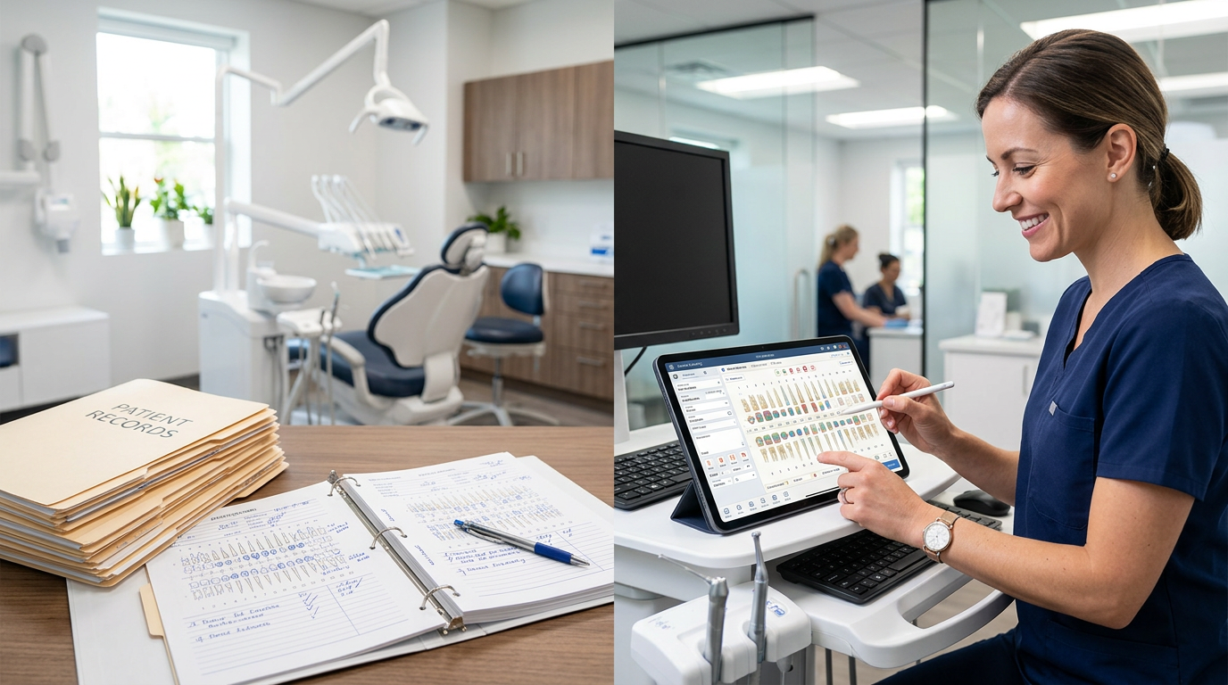
For decades, dental radiography depended on film-based X-rays, a method with real limitations. Film needed chemical processing, involved radiation exposure that was hard to control precisely, and offered limited diagnostic contrast. Digital radiography changed everything. Digital sensors, whether intraoral or extraoral, capture X-ray data directly and convert it into electronic signals that computers can process. That shift opened the door for advanced imaging software, which now acts as the brain behind every digital image.
The progress didn't stop at digital X-rays. Technologies like Cone Beam Computed Tomography (CBCT), intraoral cameras, and optical coherence tomography (OCT) followed, each generating vast amounts of data. Dental imaging software organizes, enhances, and presents that data in a clinically meaningful way. This move from analog to digital was never just about convenience, it unlocked new dimensions of diagnostic accuracy and treatment foresight.
Early Caries Detection: Unveiling the Invisible
Early detection of dental caries is paramount. The sooner you identify a carious lesion, the more conservative and less invasive the treatment, which preserves tooth structure and prevents further damage. Traditional approaches, including visual examination and conventional film radiography, often miss incipient lesions, especially those located interproximally, hidden occlusally beneath intact enamel, or in the earliest stages of demineralization.
This is exactly where advanced dental imaging software proves indispensable.
1. Enhanced Image Quality and Manipulation
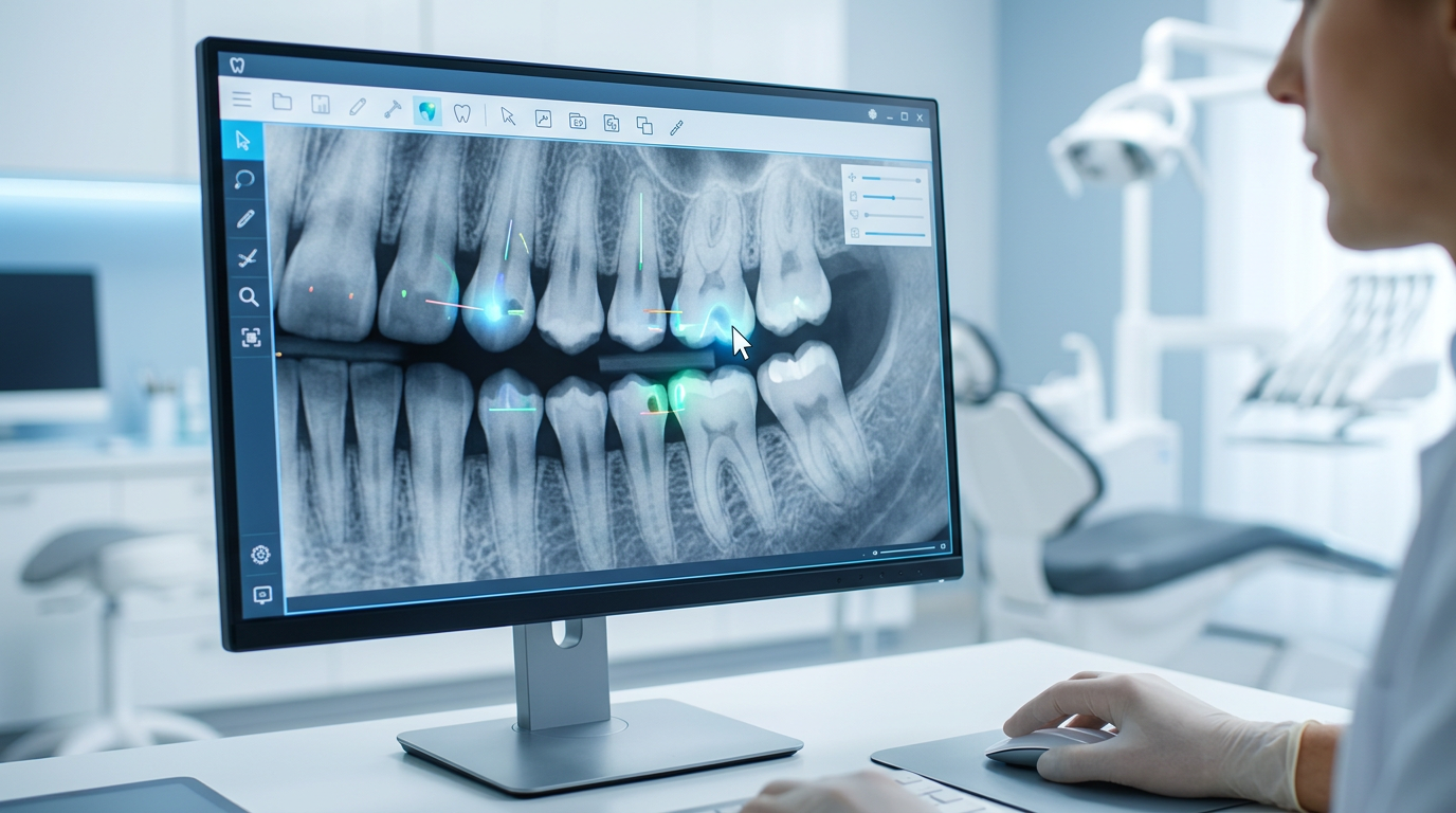
- Magnification: Software lets clinicians magnify areas of interest without losing resolution, bringing subtle demineralization into clear view.
- Contrast and Brightness Adjustment: Customizable settings optimize image contrast and brightness, highlighting subtle changes in tooth density that signal early caries.
- Sharpening Filters: Digital filters enhance the clarity of enamel and dentin structures, making early lesions stand out against healthy tissue.
2. Specialized Diagnostic Tools
- Density Analysis: Some advanced software quantifies variations in tooth density, pinpointing areas of reduced mineral content that mark early demineralization, well beyond what the human eye can perceive on a standard radiograph.
- Color Mapping and Pseudo-Coloring: Certain programs use pseudo-coloring to translate subtle greyscale differences into easy-to-read color gradients, drawing attention to potential carious lesions.
- Subtraction Radiography: This sophisticated technique superimposes and subtracts images taken at different time points. Any change in radiopacity between them, whether demineralization from caries or remineralization, is highlighted and made quantifiable, which is especially valuable for monitoring whether a lesion is progressing or arresting.
3. 3D Imaging (CBCT) for Complex Cases
- Conventional 2D radiographs handle many caries detections well, but CBCT adds a three-dimensional perspective. For complex occlusal caries, recurrent caries under restorations, or advanced lesions, CBCT reveals the depth and spread of decay in ways 2D images cannot. This volumetric data helps distinguish superficial from deeper lesions, particularly in molars where cuspal overlay obscures conventional views.
4. AI-Powered Caries Detection
- Integrating Artificial Intelligence (AI) and machine learning into dental imaging software marks a true frontier in caries detection. Trained on vast datasets of dental images, AI identifies patterns of caries with consistency and speed that often exceed human observers. These systems highlight suspicious areas, act as a 'second pair of eyes,' and help reduce diagnostic errors, particularly for incipient lesions that might otherwise be overlooked. AI doesn't replace clinician judgment, it works as a powerful diagnostic aid.
Optimizing Treatment Planning: A Blueprint for Success
Once you detect caries, effective treatment planning is the next critical step. Dental imaging software gives clinicians a robust platform to build precise, conservative, patient-specific treatment strategies.
1. Accurate Lesion Assessment
- Depth and Extent: Software measures caries depth and width precisely, helping you determine whether a lesion has reached the dentin, sits close to the pulp, or needs minimal intervention. That clarity guides the choice between remineralization therapy, a conservative filling, or a more extensive restoration.
- Relationship to Adjacent Structures: For interproximal caries, imaging software visualizes the lesion's proximity to the adjacent tooth, periodontal ligament, and alveolar bone, informing your restorative material and technique.
2. Virtual Treatment Simulation
- Restorative Planning: Some advanced software virtually reconstructs the tooth after caries removal, letting clinicians preview restoration outcomes. This helps in selecting materials, such as composite, amalgam, or an inlay/onlay, and designing the restoration's form and function.
- Endodontic Planning: For deep caries that may require root canal treatment, CBCT images processed through imaging software reveal root canal morphology, accessory canals, calcifications, and periapical pathology. This 3D understanding is critical for successful endodontic therapy, lowering procedural risk and improving long-term prognoses.
3. Patient Education and Engagement
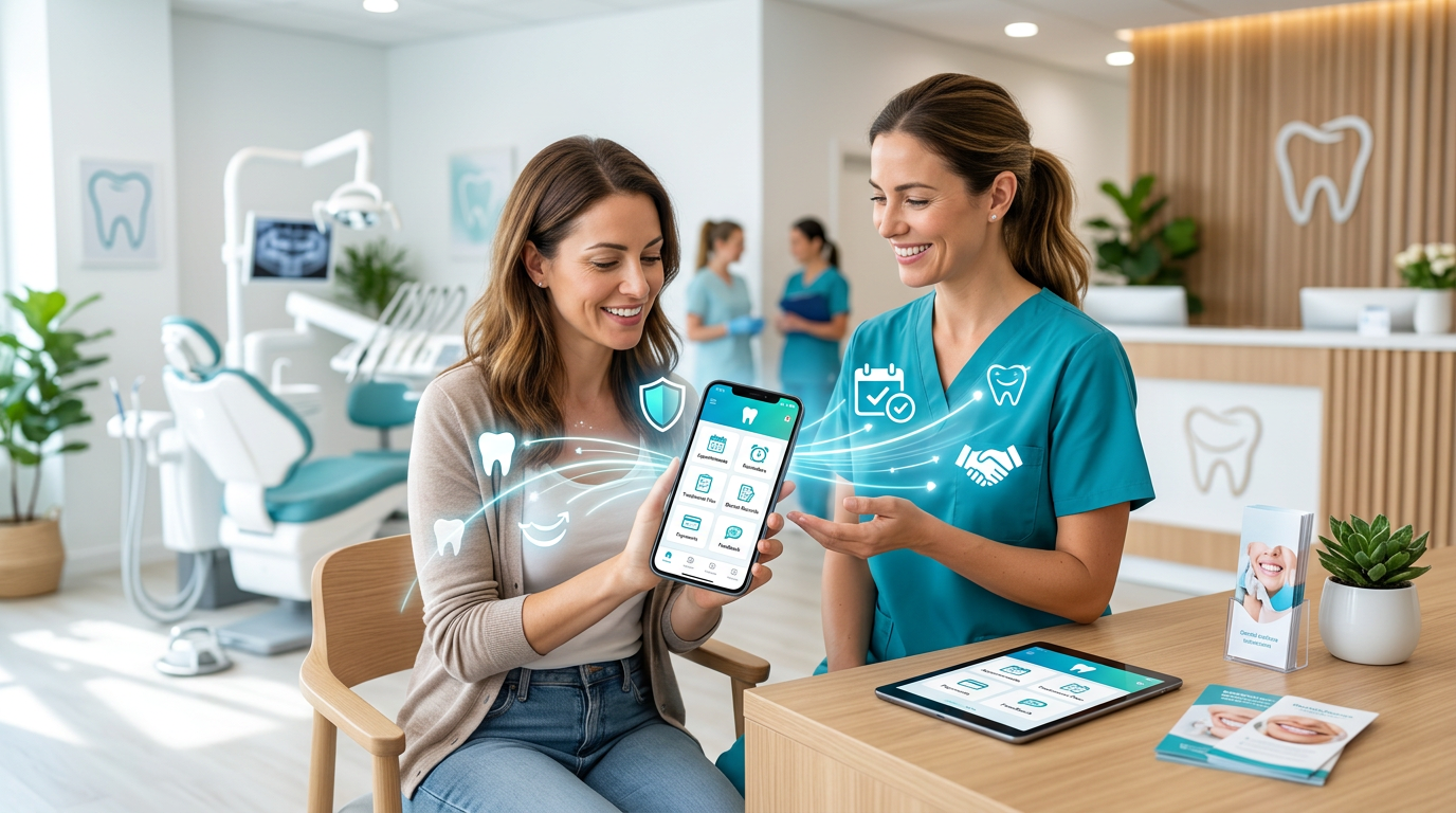
- Visual Communication: One of the most powerful uses of imaging software is patient education. By displaying high-resolution images, highlighting carious lesions, and showing animated explanations of treatment options, clinicians communicate the diagnosis and plan clearly. Patients who understand their condition are far more likely to accept and follow recommendations.
- Treatment Transparency: Clear visual aids build trust. Patients can see the 'before' and 'after' potential, grasp why intervention matters, and feel genuinely involved in their care decisions.
4. Integrated Workflow and Collaboration
- Interoperability: Modern dental imaging software often integrates with practice management systems, electronic health records (EHRs), and CAD/CAM software, creating a unified digital workflow that streamlines appointments, data, and team communication, including with external labs. A connected patient management system keeps imaging, records, and history in one place.
- Referral Management: For cases needing a specialist, such as oral surgery or orthodontics, digital images and reports can be shared securely and efficiently, ensuring coordinated care and informed decisions.
The Future Is Now: Emerging Trends in Dental Imaging Software
Dental imaging software keeps advancing, promising even more sophisticated capabilities:
- Deeper AI Integration: Expect AI to move beyond detection into predictive analytics, forecasting caries risk and suggesting personalized preventive strategies from longitudinal data.
- Augmented Reality (AR) and Virtual Reality (VR): AR and VR could let dentists superimpose digital patient data onto the patient during exams or rehearse complex procedures virtually, improving precision and training.
- Real-Time Imaging and Monitoring: Non-ionizing imaging paired with advanced software could enable continuous, real-time monitoring of oral health, offering immediate feedback on hygiene effectiveness or lesion progression.
- Enhanced Interoperability and Cloud Computing: Greater standardization and cloud platforms will make data sharing and collaboration across practices and specialties seamless, building a truly interconnected dental ecosystem by 2026 and beyond.
Challenges and Considerations
The benefits are profound, but adopting advanced dental imaging software comes with considerations:
- Cost: The initial investment in software, hardware, and training can be significant.
- Training and Adoption: Dental teams need thorough training to use the software's full capabilities.
- Data Security and Privacy: Protecting sensitive patient data in a digital environment is paramount and demands robust security protocols and regulatory compliance.
- Radiation Dose Management: Digital radiography generally reduces radiation exposure compared to film, but responsible use and the ALARA (As Low As Reasonably Achievable) principle remain critical, especially with 3D imaging.
A Paradigm Shift Toward Proactive, Personalized Care
Dental imaging software has moved dentistry from a reactive field to a proactive, precision-driven discipline. By delivering unmatched clarity in early caries detection and enabling sophisticated treatment planning, these tools help dental professionals provide care that is more accurate, more conservative, and ultimately better for patients. As AI, advanced analytics, and immersive visualization mature, imaging software will only become more central to the modern dental practice.
For Denti360, embracing these technologies isn't optional, it's essential to delivering the highest standard of oral healthcare in 2026 and beyond. The right software turns diagnostic clarity into operational efficiency across your whole practice. To see how Denti360 ties imaging insights into a connected clinic workflow, request a personalized demo today, or browse more dental technology insights on our blog.