
How Dental Imaging Software Sharpens Orthodontic Treatment Planning
Dental imaging software has made imprecise orthodontic treatment planning a thing of the past, and practices still relying on plaster models and flat X-rays are leaving precision, predictability, and case acceptance on the table. Orthodontics, the branch of dentistry focused on diagnosing, preventing, and correcting malpositioned teeth and jaws, has been transformed by digital technology. For professionals seeking to elevate their practice and deliver superior outcomes, understanding and leveraging these tools is now a necessity. This guide explores how modern dental imaging software achieves precise orthodontic treatment planning while improving efficiency, accuracy, and patient satisfaction.
The journey to a perfect smile begins with accurate diagnostics. Traditional methods, while foundational, struggled to visualize the intricate three-dimensional structures of the craniofacial complex. Analog X-rays provided planar views that left much to interpretation, and physical impressions could distort or break. Modern imaging software integrates data from intraoral scanners, CBCT machines, and digital photographs to build a comprehensive, multi-dimensional virtual model of the patient's dentition and surrounding anatomy. This digital leap streamlines diagnosis and gives orthodontists previously unattainable insight, paving the way for truly customized, effective treatment.
The Core Components of Dental Imaging Software in Orthodontics
At its heart, dental imaging software for orthodontics is a powerful suite that acquires, processes, analyzes, and visualizes patient data. Understanding its key components clarifies its impact on treatment planning.
1. Data Acquisition and Integration
The first step is gathering high-quality patient data. Modern systems integrate multiple digital sources seamlessly:
- Intraoral scanners: Capture highly accurate 3D digital impressions of teeth and gums, replacing physical molds. The digital models are more precise, eliminate storage issues, and can be shared instantly.
- Cone Beam Computed Tomography (CBCT): Provides detailed 3D volumetric images of bone, teeth, soft tissues, and air spaces. Unlike conventional 2D panoramic or cephalometric radiographs, CBCT offers a complete view of the craniofacial complex, crucial for identifying impacted teeth and assessing root angulation, bone density, and airway patency.
- Digital photography: High-resolution extraoral and intraoral photographs provide aesthetic and functional information that aids smile design and facial analysis.
- 2D radiography (cephalometric and panoramic X-rays): Still useful for specific measurements and comparisons, often integrated and traced digitally within the software.
The software acts as a central hub, consolidating these data sets into a unified patient record, a 'digital twin' of the patient's oral and maxillofacial anatomy.
2. Advanced Visualization Tools
Once data is acquired, the software's visualization capabilities let orthodontists view anatomy in:
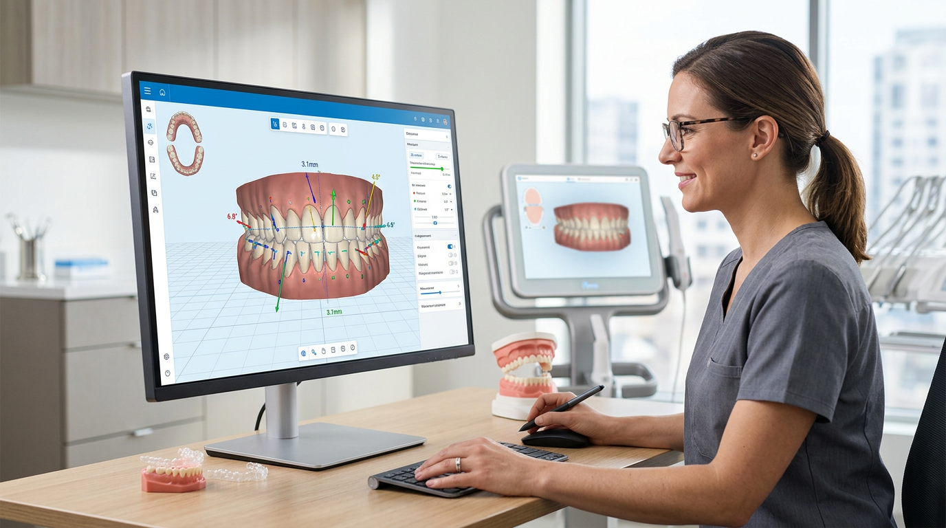
- 3D renderings: Manipulate and rotate models to view teeth, bone, and soft tissues from any angle, revealing subtleties missed in 2D.
- Cross-sectional views: Examine specific anatomical slices to assess root proximity, alveolar bone levels, and vital structures.
- Superimposition: Overlay different scans or models (e.g., pre-treatment versus post-treatment simulation, or bone versus soft tissue) to analyze changes and plan movements.
3. Diagnostic and Measurement Features
Beyond visualization, the software provides diagnostic tools:
- Cephalometric analysis: Automated tracing and analysis of cephalometric radiographs provide standard measurements for skeletal and dental relationships.
- Airway analysis: Identify and quantify airway constrictions from CBCT data, crucial for patients with potential sleep-disordered breathing.
- TMJ analysis: Evaluate temporomandibular joint morphology and position using CBCT data.
- Space analysis: Accurately measure arch length discrepancies and calculate space requirements for tooth movement.
- Impacted tooth localization: Precisely pinpoint the 3D location and relationships of impacted teeth.
4. Treatment Planning and Simulation Modules
This is where the software truly shines, allowing digital manipulation of teeth and jaws to simulate outcomes:
- Virtual tooth movement: Move teeth individually or collectively to ideal positions, simulating the effects of braces or aligners.
- Surgical planning: For orthognathic cases, simulate bone cuts and movements to visualize final occlusal and aesthetic outcomes.
- Appliance design: Design custom brackets, wires, and clear aligners from the planned movements, with many systems exporting files for 3D printing of aligner molds or indirect bonding trays.
- Occlusion analysis: Simulate and analyze intercuspal relationships and occlusal contacts in the planned final position.
Leveraging Dental Imaging Software for Greater Precision
Integrating these features profoundly improves the precision of orthodontic planning.
1. Comprehensive 3D Diagnosis and Assessment
Traditional 2D imaging suffered from superimposition and distortion. With CBCT and intraoral scans, orthodontists gain a true 3D understanding of:
- Root angulation and proximity: Essential for avoiding root resorption or collision during movement, especially near cortical bone or adjacent teeth.
- Alveolar bone volume: Critical for assessing the feasibility of expansion, intrusion, or extrusion and preventing dehiscence or fenestration.
- Impacted tooth position: Precisely locate impacted canines or third molars relative to vital structures like the inferior alveolar nerve, aiding safe surgical exposure or extraction planning.
- Skeletal discrepancies: Diagnose sagittal, transverse, and vertical skeletal imbalances more accurately than with 2D methods alone.
2. Predictable Treatment Simulation and Outcome Visualization
One of the most powerful capabilities is virtually 'treating' the patient before treatment begins, which allows for:
- Step-by-step movement planning: Plan each stage of tooth movement, predicting the exact forces and vectors required.
- Conflict identification: Spot issues like premature contacts, root collisions, or bone limitations early, allowing adjustments before treatment starts.
- Optimized appliance design: Design custom appliances tailored to the patient's anatomy and planned movements for more efficient, effective treatment.
- Patient engagement: Show patients a realistic simulation of their expected final outcome, improving case acceptance and compliance, because patients who can visualize their future smile invest more in the process.
3. Facilitating Interdisciplinary Collaboration
Orthodontic treatment often involves oral surgeons, periodontists, and restorative dentists. Dental imaging software enhances collaboration by:
- Seamless data sharing: Digital files are shared easily and securely, eliminating physical models or films.
- Unified treatment plans: All specialists view and contribute to a single comprehensive digital plan, ensuring coordination for complex cases involving orthognathic surgery or prosthodontics.
- Pre-surgical planning: For orthognathic cases, surgeons plan bone cuts and movements precisely, often with custom surgical guides printed from the digital plan.
4. Streamlining Workflow and Enhancing Efficiency
Beyond clinical precision, the software improves practice management:
- Reduced chair time: More accurate planning means fewer adjustments, fewer appointments, and shorter overall treatment times.
- Elimination of physical models: Digital impressions remove the need for plaster models, saving storage space, reducing material costs, and preventing model damage.
- Automated processes: Features such as cephalometric tracing and volumetric measurements are automated, saving significant time versus manual methods.
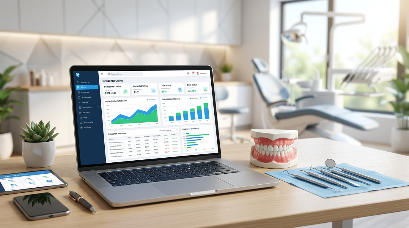
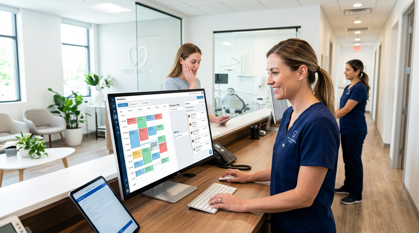
- Integration with practice management systems: Many solutions integrate with practice management platforms, creating a cohesive digital ecosystem for records, scheduling, and billing. Connecting imaging to streamlined patient management keeps every record in sync.
5. Error Reduction and Risk Mitigation
The detailed insights from dental imaging software help minimize treatment risks:
- Avoiding nerve damage: Precise localization of the inferior alveolar nerve in CBCT scans prevents iatrogenic injury during extractions or implant placement in orthodontically prepared sites.
- Preventing root resorption: Visualizing root proximity and angulation lets orthodontists plan movements that minimize stress on roots, reducing resorption risk.
- Optimizing anchorage: Accurately assess bone quality and quantity for temporary anchorage devices (TADs), ensuring stable anchorage and predictable movement.
The Future of Orthodontic Treatment Planning: AI and Beyond
Dental imaging software keeps evolving. Looking ahead to 2025 and beyond, artificial intelligence (AI) is poised to further revolutionize orthodontic planning. AI algorithms are already being developed to:
- Automate diagnosis: Identify anomalies, classify malocclusions, and perform cephalometric analyses with greater speed and accuracy.
- Predict treatment outcomes: Drawing on vast datasets of successful cases, AI could predict the most efficient pathways and potential challenges for individual patients.
- Personalize treatment recommendations: Suggest highly customized plans, including appliance choices and force systems, tailored to unique patient biologies.
- Monitor progress in real time: AI-powered tools could analyze intra-treatment scans to assess progress and recommend adjustments, further optimizing the trajectory.
Virtual reality (VR) and augmented reality (AR) also hold promise, letting orthodontists immerse themselves in a patient's digital anatomy for more intuitive planning and even remote consultations. Cloud-based platforms will continue to enable secure data storage, accessibility, and collaboration from anywhere in the world.
Embrace the Digital Edge in Orthodontics with Denti360
For Denti360, a leader in dental healthcare and technology, championing advanced dental imaging software in orthodontic practices is paramount. The precision, efficiency, and enhanced patient experience these tools deliver represent a significant leap forward in oral healthcare. By providing orthodontists with detailed 3D insights, predictive simulations, and streamlined workflows, this technology empowers them to deliver superior outcomes with greater confidence. The transition to a fully digital orthodontic practice is an investment with undeniable returns in clinical excellence, operational efficiency, and patient satisfaction, and it sets a new standard for care. Explore the Denti360 platform features or contact our team to unlock a future of unparalleled precision in orthodontics.