
Optimizing Early Caries Detection With Dental Imaging Software
Dental imaging software gives your practice its best shot at catching interproximal caries before they ever become a cavity. These "hidden" lesions sit between the teeth, where direct vision and tactile instruments fail, and by the time they are clinically obvious they often demand far more extensive restorative work. Advanced imaging software answers this diagnostic gap with precision, enhanced visualization, and data-driven insights that are transforming early caries detection. For Denti360, a leader in dental healthcare and technology, leveraging these innovations is key to elevating patient care.
Interproximal caries pose a real diagnostic challenge because their location resists visual and tactile examination. Left undetected, initial enamel lesions advance through the dentin, leading to cavitation, pain, and potential tooth loss. Integrating sophisticated dental imaging software into daily practice addresses these challenges and empowers clinicians to make informed decisions, intervene early, and improve long-term oral health outcomes.
Why Early Caries Detection Matters
Dental caries progression typically starts with demineralization of the enamel surface. Interproximal surfaces are especially susceptible to plaque accumulation and difficult to clean effectively. If left untreated, these initial enamel lesions advance through the dentin, leading to cavitation, pain, and potentially tooth loss. Modern dentistry increasingly emphasizes prevention and minimal intervention, making early detection of non-cavitated or incipient lesions critical.
Early detection enables preventive strategies such as fluoride application, sealants, or remineralization therapies that can halt or even reverse the carious process before irreversible damage occurs. This preserves tooth structure and reduces the need for complex, costly, time-consuming restorations. For patients, that means less discomfort, fewer appointments, and a more positive experience. For practices, it signals a more proactive, patient-centric, and economically sustainable model of care.
Limitations of Traditional Interproximal Caries Detection
Before examining modern software, it helps to acknowledge the limits of conventional methods:
- Visual Examination: Essential, but direct inspection of interproximal surfaces is hindered by tooth contact points and soft tissue, making early, non-cavitated lesions almost impossible to spot.
- Tactile Examination (Explorer): A dental explorer can detect surface irregularities and softened enamel, but its efficacy interproximally is limited, and aggressive use risks iatrogenic damage by cavitating a demineralized but non-cavitated surface.
- Conventional Radiography (Bitewings): Long the gold standard for interproximal caries, bitewings present several challenges:
- 2D Representation: Radiographs project 3D structures in 2D, causing superimposition and potential misinterpretation, and lesions can be obscured by overlying tooth structure or restorations.
- Threshold for Detection: Caries must reach a certain size and depth to appear radiographically; demineralization typically needs to affect 30-50% of the enamel thickness before it is detectable on a radiograph.
- Image Quality Variability: Exposure settings, sensor type, and processing significantly affect image quality and diagnostic accuracy.
- Radiation Exposure: Though minimized, radiation remains a consideration for routine or frequent imaging.
These limitations underscore the need for more sensitive, precise tools that overcome the challenges of traditional methods and improve diagnostic accuracy and treatment planning.
How Dental Imaging Software Transforms Detection
A cornerstone of digital dentistry, dental imaging software uses advanced algorithms and processing to enhance the diagnostic utility of imaging modalities. It is not merely a viewer; it is a powerful analytical tool that extracts, interprets, and presents data in ways analog methods cannot. Here is how it transforms interproximal caries detection.
1. Enhanced Visualization and Image Processing
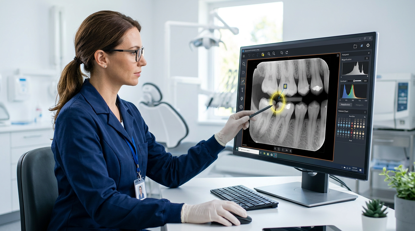
Modern software offers tools that optimize image quality and surface subtle details:
- Contrast and Brightness Adjustments: Clinicians manipulate parameters in real time to accentuate density changes that indicate demineralization, making early lesions more discernible than on raw images.
- Magnification: High-resolution images magnify extensively without pixelation for close examination of critical areas.
- Filtering and Sharpening: Filters reduce noise, sharpen edges, and enhance visibility of subtle enamel and dentin changes, with some software offering dedicated caries-detection filters.
- Color Mapping and Pseudocoloring: Advanced software applies color to grayscale images based on pixel density, making demineralization stand out more clearly.
2. Quantitative Analysis and Measurement
Beyond qualitative assessment, imaging software adds quantitative capabilities:
- Lesion Depth Measurement: Advanced tools precisely measure lesion depth and width, providing objective data on progression, useful for monitoring incipient lesions over time.
- Density Analysis: By analyzing pixel density, software identifies subtle demineralization before it is overtly visible, offering an objective assessment of tissue density changes.
- Comparative Analysis: Software overlays current images with previous ones to track changes in lesion size or density over time, invaluable for deciding whether to intervene or continue monitoring.
3. Integration With Advanced Imaging Modalities
The true power of imaging software emerges when it integrates with cutting-edge technologies:
- Digital Radiography (Sensors and PSPs): High-resolution digital sensors deliver immediate images with a wider dynamic range than film, enabling greater post-processing. The software manages these images, reduces radiation, and enhances diagnostic yield.
- Intraoral Scanners (for Surface Analysis): Primarily used for restorative dentistry, some scanners and their software are developing the ability to detect early surface demineralization by analyzing changes in light reflection or using fluorescence technology.
- Cone Beam Computed Tomography (CBCT): Generally not indicated for routine caries detection due to higher radiation and scatter artifacts, but specialized CBCT systems with very small fields of view and high resolution can offer insights into complex cases difficult to assess on 2D radiographs, with software enabling multi-planar reconstruction and 3D visualization.
- Fluorescence-Based Caries Detection Systems: These use specific wavelengths of light to excite porphyrins produced by cariogenic bacteria, causing them to fluoresce red while healthy tooth structure fluoresces green. Integrated software quantifies these signals to detect early demineralization and monitor remineralization, with examples including DIAGNOdent and the Canary System, processing raw fluorescence data into visual and numerical outputs.
4. Artificial Intelligence and Machine Learning
AI and machine learning represent the frontier of imaging software and are poised to revolutionize caries detection:
- Automated Caries Detection: AI algorithms trained on vast image datasets automatically identify potential lesions with high accuracy, highlighting suspicious areas that warrant closer examination.
- Risk Assessment: Beyond detection, AI analyzes patient history, demographics, and imaging data to predict caries risk and guide personalized preventive strategies.
- Decision Support: AI acts as a sophisticated second opinion, cross-referencing findings with established criteria and suggesting diagnoses, reducing variability and improving consistency.
- Workflow Efficiency: By automating parts of the diagnostic process, AI streamlines workflows so dentists can focus more on patient interaction and treatment planning.
As of 2025, several dental software companies are integrating AI modules that promise to significantly enhance the sensitivity and specificity of caries detection, particularly for early interproximal lesions.
Best Practices for Implementing Dental Imaging Software
To fully leverage imaging software for early interproximal caries detection, practices should follow these best practices:
- High-Quality Digital Radiography: Invest in high-resolution sensors or phosphor plates and apply consistent radiographic technique for reproducible, diagnostically valuable images.
- Staff Training: Ensure dentists, hygienists, and assistants are thoroughly trained in the software, its features, and recognizing subtle radiographic signs of caries.
- Regular Calibration and Updates: Keep software current for new features, improved algorithms, and fixes, and calibrate imaging equipment regularly for consistent performance.
- Integrated Workflow: Choose software that integrates with your practice management system and other diagnostic tools, such as intraoral cameras and fluorescence devices, for efficiency and data consistency, supported by streamlined patient management.
- Patient Education: Use enhanced visualization to educate patients, since magnified images with highlighted lesions improve understanding and acceptance of treatment.
- Documentation: Maintain meticulous digital records, since easy storage, retrieval, and comparison over time is a major advantage of digital imaging.
- Combine Modalities: No single tool is perfect, so combine imaging software with clinical examination, fluorescence technology, and patient history for comprehensive diagnosis.
The Denti360 Advantage: Pushing the Boundaries of Caries Detection
Denti360 is at the forefront of integrating cutting-edge dental imaging software and AI into practice. Our commitment is to give professionals tools that exceed the demands of modern diagnostics. Our solutions are designed to:
- Enhance Diagnostic Accuracy: Crystal-clear images, advanced processing, and AI-powered insights help identify interproximal caries at their earliest, most treatable stages.
- Improve Patient Outcomes: Early detection means less invasive treatment, better preservation of natural tooth structure, and fewer long-term dental problems.
- Increase Practice Efficiency: Streamlined workflows, automated analysis, and seamless integration reduce diagnostic time and improve operations.
- Facilitate Patient Communication: Intuitive visualization helps dentists explain findings clearly, fostering trust and improving treatment acceptance.
- Support Continuous Learning: Our platforms offer case review, comparison, and integration with educational resources to support professional development.
As dental technology advances, Denti360 remains dedicated to research and development, ensuring clients have access to the most innovative and reliable tools for optimizing patient care in 2025 and beyond.
Detect Caries Earlier With Denti360
The move from traditional film radiography to sophisticated dental imaging software is a significant leap toward optimal oral health, and for elusive interproximal caries it is revolutionary. Enhanced visualization, quantitative analysis, and the growing power of artificial intelligence empower a proactive approach to dental care, turning early detection into an achievable standard rather than an aspiration. Practices that embrace these technologies elevate diagnostic precision and treatment efficacy while positioning themselves as leaders in minimally invasive care. See how Denti360 brings these capabilities together by exploring our platform features, or contact our team to optimize your caries detection workflow today.