
Early caries detection is one of the most consequential decisions a dentist makes every day. Catch a lesion before it breaches the enamel and you can arrest it with remineralisation; miss it and your patient faces a restoration, or worse, a root canal. For decades, the trusty dental X-ray has been the frontline tool for this call. Now, AI-powered dental imaging is reshaping that picture — and every dental practice in India needs to understand what this shift means for patient outcomes and clinical workflow.
How Traditional Radiographs Work — and Where They Fall Short
Bitewing and periapical radiographs produce two-dimensional grayscale images by recording the differential absorption of X-rays through tooth and bone. Dense, mineralised enamel appears radiopaque; areas of demineralisation appear darker. In skilled hands, this technique has guided dental diagnostics for over a century, and it remains an affordable, widely available standard of care across Indian clinics.
The limitation, however, is physical. Research consistently shows that conventional radiographs cannot reliably detect interproximal lesions until roughly 30–40% of the enamel mineral has already been lost. By that point, the window for non-invasive remineralisation therapy has often closed. Add to that the variability between practitioners — different levels of experience, visual fatigue, inconsistent viewing conditions — and it becomes clear that the human eye acting alone is not always a reliable detector of early caries.
- Two-dimensional projection: Overlapping structures can mask early lesions, especially in the proximal surfaces of posterior teeth.
- Detection threshold: Significant mineral loss must occur before a lesion is visible, delaying the opportunity for preventive intervention.
- Inter-observer variability: Studies show meaningful differences in diagnosis between clinicians reviewing the same image, affecting consistency of care.
- Limited depth information: Traditional X-rays cannot convey the three-dimensional extent of a lesion, complicating treatment planning for borderline cases.
What AI-Powered Dental Imaging Actually Does
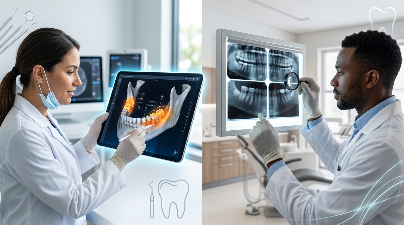
AI in dental imaging is not a separate scanner or X-ray machine — it is an analytical layer that sits on top of your existing digital radiography workflow. Deep-learning algorithms, trained on hundreds of thousands of annotated radiographic images reviewed by expert clinicians, learn to recognise the subtle pixel-level patterns that correspond to early demineralisation, cavitation, and interproximal lesions. When you capture a standard bitewing, the AI overlay highlights regions of concern in seconds, giving the clinician a flagged image alongside their own unassisted view.
Several peer-reviewed studies published in journals such as Dentomaxillofacial Radiology and the Journal of Dental Research have reported that AI-assisted caries detection achieves higher sensitivity for early-stage lesions compared to unassisted human reading of the same radiographs. Crucially, the AI does not fatigue, does not have a bad day, and applies identical criteria to every image — eliminating much of the inter-observer variability that clouds traditional diagnosis.
- Automated feature detection: Identifies subtle density changes in enamel and early dentin that are below the threshold of reliable human detection.
- Consistent, objective analysis: The same algorithm assesses every radiograph the same way, regardless of clinic volume or time of day.
- Speed: Flagging takes seconds, keeping your chairside workflow moving without adding consultation time.
- Longitudinal monitoring: AI can compare current radiographs to archived images and quantify lesion progression or arrest — invaluable for minimal intervention dentistry protocols.
- Patient communication: A colour-coded overlay on a digital image is far easier to explain to a patient than pointing at a faint grey shadow, improving case acceptance for preventive treatment.
AI vs. Traditional Radiographs: A Direct Comparison for Indian Dental Practices
When evaluating any new technology for a dental practice in India, the questions are practical: Does it improve outcomes? Does it fit the workflow? Is the investment justified? Here is how AI-assisted imaging measures up against standalone traditional radiography across the criteria that matter most.
Diagnostic Accuracy
AI-assisted systems consistently demonstrate superior sensitivity for incipient and early-enamel lesions — the very lesions where early intervention makes the biggest clinical difference. Traditional radiographs remain strong for detecting moderate-to-advanced lesions, but that is already late-stage detection. For a preventive dentistry model, AI adds genuine diagnostic value.
Consistency and Standardisation
In a busy multi-dentist clinic or a franchise dental chain, ensuring diagnostic consistency across all practitioners is a real challenge. AI provides a standardised analytical benchmark that is not affected by individual practitioner variability, supporting quality assurance and peer review processes without additional administrative burden.
Workflow Integration
Modern AI dental imaging tools integrate directly with digital radiography sensors and dental practice management software like Denti360, meaning there is no separate login, no additional hardware, and no disruption to existing processes. The radiograph is captured as usual; the AI annotation appears alongside it in the patient record within moments.
Patient Experience and Trust
Patients increasingly expect their dental clinic to use visible technology. Showing a patient a flagged, colour-annotated radiograph and explaining the finding in plain language — supported by AI analysis — builds confidence in the diagnosis and the recommendation, which directly improves treatment acceptance rates.
The Right Way to Think About AI in Your Practice
AI-powered dental imaging is not a replacement for clinical judgement — it is a decision-support tool. The dentist remains responsible for synthesising the radiographic evidence with clinical examination findings, patient history, dietary habits, saliva assessments, and patient preferences. What AI does is raise the floor of diagnostic sensitivity so that fewer early lesions are missed, and it provides an objective, documented second opinion that is especially valuable in complex or ambiguous cases.
This human-AI collaboration is where the real gains lie. Practices that treat AI as a background quality-check rather than an automated decision-maker will get the most out of it — and their patients will benefit the most.
For Indian dental clinics looking to build a preventive care model, differentiate their services, and reduce the incidence of late-stage restorative treatments, AI-assisted imaging is a meaningful and practical step forward. Dental software India-wide is evolving rapidly, and integrated dental practice management platforms are already making these tools accessible to clinics of all sizes.
Conclusion
Traditional radiographs laid the foundation of modern dental diagnostics, and they are not going away. But when it comes specifically to early caries detection — where the stakes for patient outcomes are highest — AI-powered imaging offers measurable advantages in sensitivity, consistency, and clinical communication that standalone X-ray interpretation cannot match. The combination of both, embedded in a robust dental practice management workflow, represents the current gold standard.
If you want to see how AI-assisted imaging integrates with patient records, appointment scheduling, and treatment planning in a single platform, book a free Denti360 demo today and experience what modern dental practice management looks like in practice.