
How Dental Charting Software Sharpens Caries Detection Accuracy
Dental charting software has become essential for catching caries you might otherwise miss, the early lesions hidden in challenging anatomy that drive misdiagnosis and unnecessary intervention. Caries, commonly known as tooth decay, remains one of the most prevalent chronic diseases globally, and early, accurate detection is paramount to successful prevention, minimally invasive treatment, and preserving tooth structure. Traditional detection relied on visual inspection, tactile examination with an explorer, and conventional two-dimensional radiography. Those methods served dentistry for decades, but their limitations often led to missed early lesions or overtreatment from misinterpretation.
Modern technology continually reshapes clinical workflows and diagnostic capabilities, and digital charting software has emerged as a transformative tool. Far beyond replacing paper charts, these systems integrate patient data, imaging, and analytical features into a comprehensive digital ecosystem. For Denti360, a leader in dental healthcare and technology, leveraging these advancements is key to giving dental professionals superior diagnostic accuracy.
This post explores five pivotal ways charting software improves caries detection: enhancing visualization, integrating data, providing analytical tools, streamlining comparison, and supporting collaborative diagnosis. Together they show why these systems are indispensable in the contemporary practice.
1. Enhanced Visualization and Magnification for Subtle Lesions
One immediate benefit of charting software is enhanced visualization and magnification. Traditional methods limit a clinician's view to what is directly visible or shown on a small film. Digital charting revolutionizes that perspective:
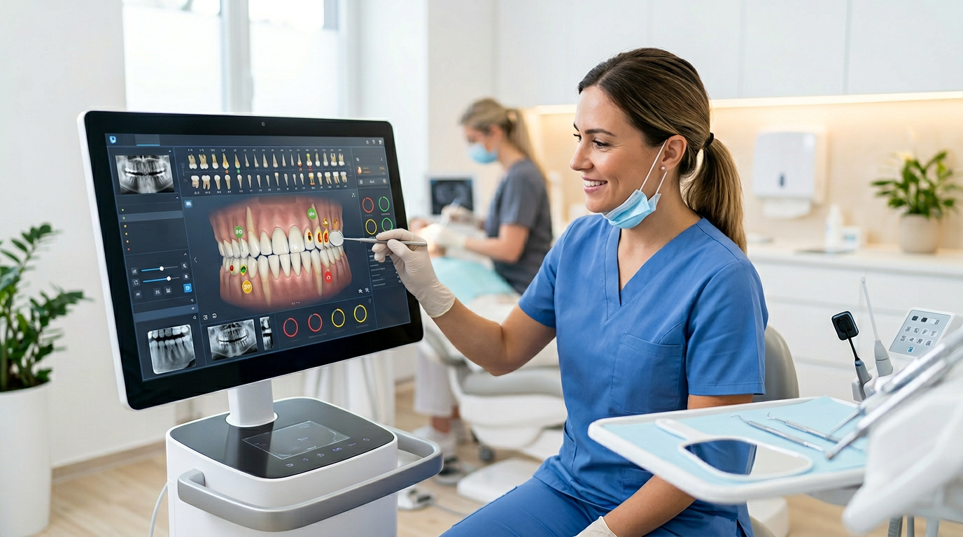
- High-resolution imaging integration: Charting software integrates with high-resolution intraoral cameras and digital radiography. Clinicians view magnified, crystal-clear images of tooth surfaces, proximal areas, and underlying bone on a large monitor, revealing subtle demineralization, faint color changes, and minute texture alterations imperceptible to the naked eye or a conventional X-ray.
- Adjustable contrast and brightness: Digital images are not fixed. Clinicians adjust contrast, brightness, and filters to highlight areas of interest. Adjusting contrast can make early enamel demineralization, or white spot lesions, more apparent, while altering brightness reveals subtle shadows of dentin caries beneath seemingly intact enamel.
- Three-dimensional views with CBCT: Systems that integrate with Cone Beam Computed Tomography offer 3D views. For recurrent caries under restorations or deeply situated lesions, a 3D perspective accurately delineates the extent and depth of decay, information 2D radiographs cannot provide, preventing diagnostic ambiguity.
- Standardized image capture: Many systems guide the user to capture images consistently, so comparison over time stays reliable. Combined with the ability to zoom and manipulate images, this makes subtle changes in tooth structure that signal early caries much more apparent.
The power to see more clearly, in greater detail, and with customizable viewing parameters significantly reduces the likelihood of missing incipient carious lesions, enabling timely intervention or non-invasive management before the disease advances.
2. Comprehensive Data Integration and Cross-Referencing
Powerful charting software acts as a central hub for all patient data. This integration and cross-referencing of diagnostic inputs is critical to detection accuracy. Instead of fragmented information across physical files or disparate systems, everything is consolidated:
- Unified patient record: The software combines clinical notes, periodontal charts, treatment history, restorative records, complaints, and risk assessments into one accessible file. Evaluating a lesion, a clinician instantly reviews past restorations, previous radiographs of the same tooth, documented sensitivity, and the caries risk profile. This kind of streamlined patient record management keeps every detail at hand.
- Correlation of intraoral images with radiographs: One of the most significant advantages is viewing intraoral photographs, digital X-rays (bitewings, periapicals, panoramic), and 3D scans side-by-side or overlaid. A suspected shadow on a bitewing correlates immediately with an intraoral camera exam, and a suspicious pit or fissure observed clinically can be confirmed or ruled out for underlying dentin involvement. This multi-modal correlation reduces guesswork and enhances diagnostic confidence.
- Tracking caries risk factors: Risk-assessment modules integrate diet, fluoride exposure, salivary flow, medical history such as xerostomia-causing medications, and past caries experience to flag higher-risk patients. This prompts clinicians to be particularly vigilant, perhaps recommending more frequent recalls or adjunctive diagnostic aids, improving the chances of early detection.
- Historical data comparison: Instantly pulling previous examinations, notes, and images from years past, such as 2025 or earlier, gives a longitudinal view. A lesion that looks benign on a single examination may prove progressive when compared against images and notes from six months or a year prior, distinguishing active lesions from arrested ones.
This holistic view, facilitated by robust data integration, ensures diagnostic decisions rest on a complete and coherent understanding of the patient's oral health status, significantly improving the accuracy of caries detection.
3. Analytical Tools and AI-Powered Assistance
The frontier of dental technology increasingly involves analytical tools and artificial intelligence embedded in charting software. These features move beyond storage to provide true diagnostic assistance:
- AI for radiographic interpretation: Some cutting-edge systems incorporate AI algorithms trained on vast radiograph datasets. These tools highlight suspicious areas such as subtle radiolucencies indicative of caries and even estimate depth or proximity to the pulp. AI supplements, rather than replaces, clinical judgment, acting as a second pair of eyes that catches lesions a fatigued or cognitively biased human eye might overlook and alerting the clinician to areas requiring closer inspection.
- Measurement and annotation tools: Software provides tools to measure lesion size, track growth, and annotate specific areas directly on images, a precision difficult with traditional methods. Measuring change in a demineralized area over time provides objective data on caries progression or arrest, aiding active surveillance versus intervention decisions.
- Color mapping and density analysis: Certain software analyzes image data to create color maps based on tissue density or other parameters, making areas of demineralization or structural change more visually prominent and transforming raw image data into easily interpretable visual cues.
- Predictive analytics: Still in nascent stages, future iterations may use a patient's historical data, genetics, and lifestyle factors to predict future caries risk or the likely progression of existing lesions, allowing even earlier intervention strategies.
By augmenting human expertise with data-driven insights and AI assistance, charting software elevates accuracy, particularly for early, ambiguous lesions.
4. Streamlined Longitudinal Comparison and Progress Tracking
Digital charting excels at streamlined longitudinal comparison and progress tracking. Caries is a dynamic process, and understanding progression or regression over time is fundamental to accurate diagnosis and management:
- Side-by-side image comparison: With a few clicks, clinicians display multiple radiographs or intraoral images of the same tooth from different appointments, such as a bitewing from 2025 next to one from six months prior. This immediate, high-resolution comparison clearly identifies subtle changes in radiolucency or surface integrity that signal caries initiation or progression, far more precise than holding physical films up to a light source.
- Automated measurement tracking: When measurements of lesion size or depth were taken at previous appointments, the software stores and displays this data so clinicians can objectively track change, invaluable for monitoring incipient lesions managed non-invasively and providing concrete evidence of whether a lesion is arresting, progressing, or regressing.
- Visual caries mapping: Many systems let clinicians graphically mark and color-code lesions directly on a digital tooth chart, providing an at-a-glance overview of a patient's caries history and current status that updates dynamically across appointments.
- Pattern recognition over time: Aggregating data across multiple visits helps recognize patterns. If a patient consistently develops interproximal caries on specific teeth despite good home care, it can prompt investigation into dietary habits or anatomical factors that might otherwise be overlooked.
- Enhanced patient education: Showing patients their own historical images and highlighting changes makes education significantly more impactful. When patients see decay progress on their own teeth over time, they are far more likely to understand the diagnosis and adhere to treatment and preventive measures.
This systematic tracking transforms diagnosis from a static snapshot into a dynamic, evidence-based process that fosters earlier detection and personalized care.
5. Facilitation of Collaborative Diagnosis and Peer Review
Modern practice benefits from collaboration, especially in complex or ambiguous cases. Charting software provides a unified, accessible platform for peer review and consultation that improves accuracy:
- Instant secure sharing of patient data: Instead of printing X-rays or photocopying notes, clinicians securely share an entire record, including high-resolution images, radiographs, clinical notes, and treatment plans, with colleagues, specialists, or for expert second opinions, ensuring consultations rest on complete and current information.
- Standardized terminology and documentation: Systems often enforce standardized terminology and documentation prompts. This consistency reduces ambiguity so that when multiple clinicians review a case, they interpret information within the same framework, leading to more accurate shared diagnoses.
- Remote consultation capabilities: For multi-location practices or specialists providing remote consultations, a general dentist easily sends a case to an endodontist or oral surgeon with all relevant diagnostic images and notes attached, leading to more informed and timely recommendations for potential pulpal involvement related to deep caries.
- Training and education tool: For dental students or new associates, robust charting software is an excellent training tool. Experienced clinicians review cases, point out subtle diagnostic features, and guide the process using real patient data, sharpening the skills of future practitioners.
- Audit trails and accountability: Systems record who accessed a file, what changed, and when, enhancing accountability with secure role-based access controls and a clear history valuable for quality assurance.
By breaking down barriers to information sharing, charting software harnesses collective expertise for more confident, accurate detection in challenging cases.
Denti360's Commitment to Precision Diagnostics
Moving from paper charting to sophisticated digital charting software represents a significant leap in caries detection. As we explored, these systems transform how professionals identify, monitor, and manage decay through enhanced visualization, comprehensive data integration, analytical assistance, longitudinal comparison, and collaborative features.
For Denti360, empowering practices with advanced, user-friendly technology is at the core of our mission. Accurate detection is not merely finding a hole in a tooth; it is intercepting disease early, preventing extensive damage, guiding minimally invasive treatment, and safeguarding patients' oral and systemic health for years to come. High-resolution imaging, AI-powered insights, and seamless data management in one platform let clinicians move from subjective assessment to objective, data-driven diagnostics. Ready to elevate your diagnostic accuracy? Connect with the Denti360 team to see our charting platform in action and bring precision diagnostics to your practice.