
Early caries detection is one of the most clinically important — and technically demanding — tasks in everyday dental practice. A lesion caught at the enamel-demineralisation stage can often be reversed with fluoride and dietary guidance. The same lesion missed for another six months may need a full restoration. The imaging technology you use in your clinic directly influences how often you catch decay early enough to act conservatively. This article compares digital dental imaging and conventional film X-rays on the criteria that matter most for early caries detection, and explains what that difference means for your patients and your practice.
How Each Technology Works
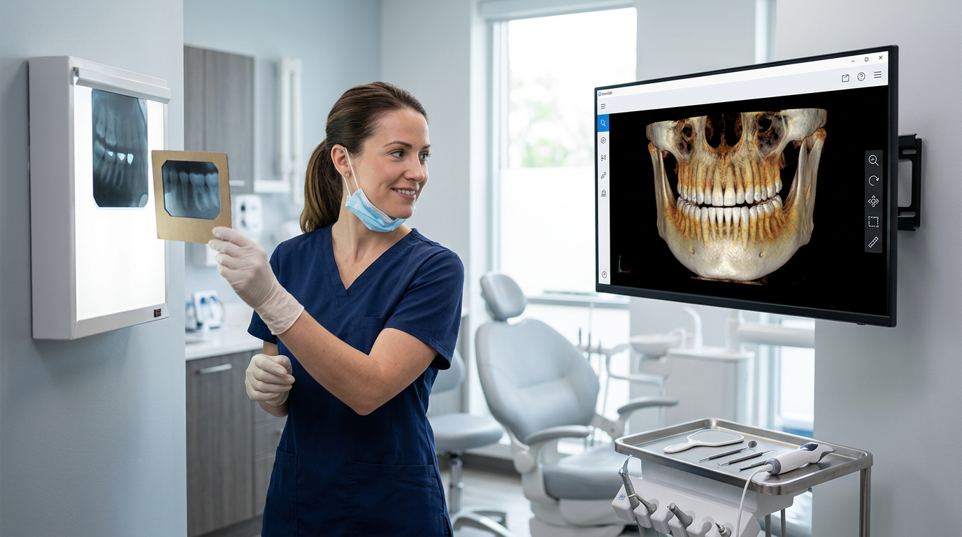
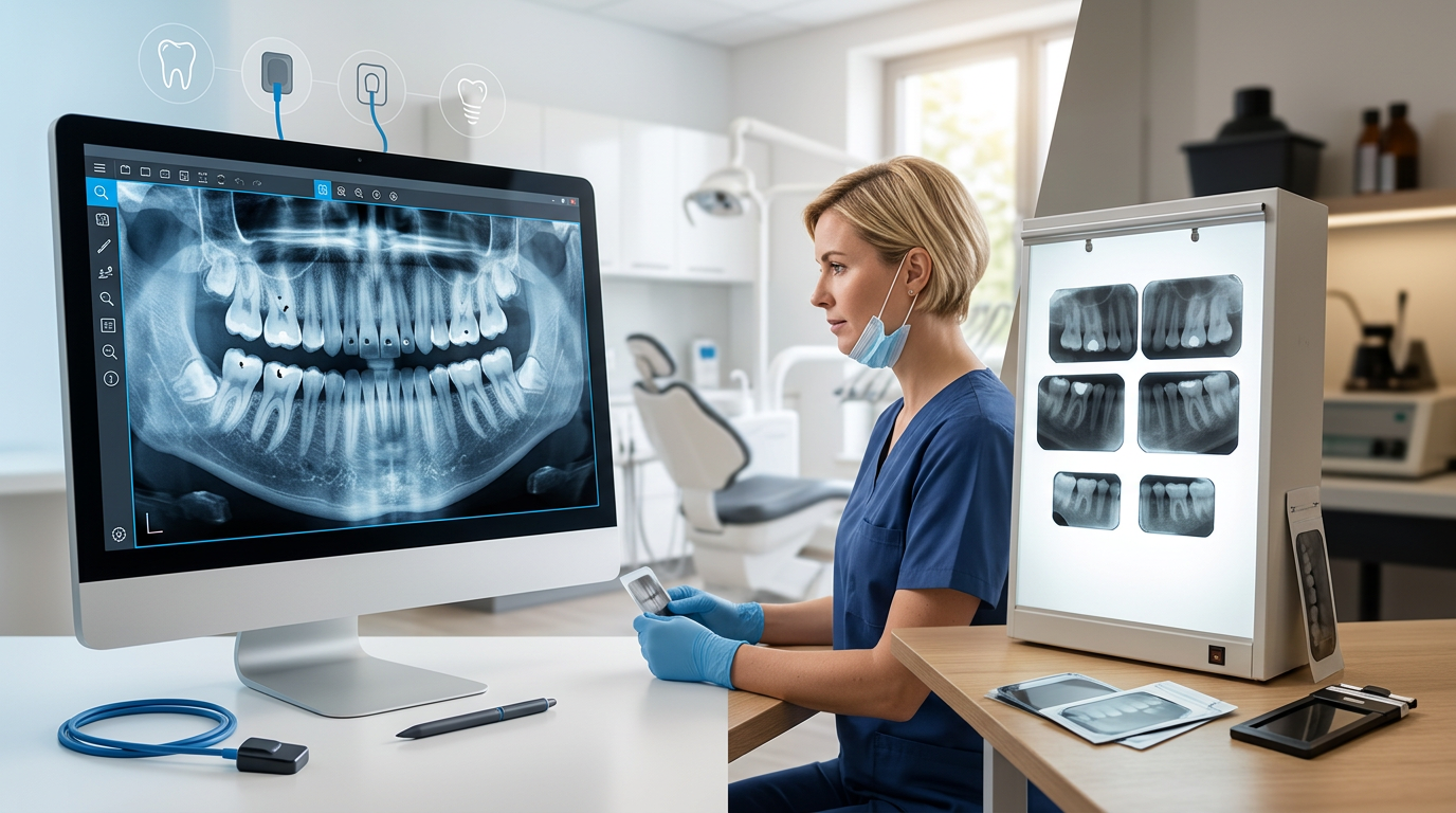
Film X-rays have been a diagnostic staple for over a century. Radiation passes through oral structures and exposes photographic film, which is then chemically developed in a darkroom to produce a static black-and-white image. Denser structures appear lighter (radiopaque); less dense or demineralised areas appear darker (radiolucent). The final image cannot be adjusted once developed.
Digital dental imaging replaces the film packet with an electronic sensor — either a rigid intraoral sensor (CCD or CMOS) or a flexible photostimulable phosphor (PSP) plate. The sensor converts X-ray energy into an electronic signal that is processed and displayed on a monitor within seconds. Unlike film, the resulting digital image can be zoomed, brightness-adjusted, contrast-enhanced, and filtered without any loss of original data.
The Early Caries Detection Gap Between Film and Digital
Incipient caries — particularly early proximal lesions that have not yet cavitated — typically manifest as faint, subtle radiolucencies. Detecting them requires both fine spatial resolution and the ability to manipulate the image to make those faint changes visible. This is precisely where film consistently underperforms compared to digital.
Resolution and Detail
Modern intraoral digital sensors capture images at resolutions that reveal fine structural detail of enamel rods and dentinal tubules. Film grain, by contrast, limits the rendition of subtle demineralisation, especially on proximal surfaces where early interproximal lesions are most common. Peer-reviewed literature has repeatedly shown that digital radiography detects small non-cavitated lesions at significantly higher rates than film — particularly for lesions confined to the outer half of enamel.
Post-Processing: The Clinical Game-Changer
The most important practical advantage of digital imaging is not raw resolution — it is the ability to process the image after capture. A clinician can:
- Increase contrast to sharpen the boundary between healthy enamel and early demineralisation
- Zoom in on a suspicious proximal contact without pixelation or loss of detail
- Apply edge-enhancement filters that highlight density transitions
- Invert the image to a colour map that makes subtle radiolucencies more visually distinct
None of these tools exist for film. A static developed image is what it is. If the contrast was suboptimal at exposure, diagnostic information is permanently lost.
Instant Retakes and Quality Control
Film X-rays require the patient to return for a separate appointment or wait for development before a clinician realises that the image was suboptimal due to positioning or exposure error. With digital imaging, the image appears on screen within seconds. If a proximal contact is overlapping and obscuring a suspected lesion area, the sensor can be repositioned immediately and a second exposure taken — with minimal additional radiation — before the patient even leaves the chair.
Radiation Dose: A Significant Patient Safety Advantage
Digital sensors are substantially more sensitive to X-ray photons than photographic film. This sensitivity means that diagnostic-quality images can be achieved with significantly lower radiation doses — often cited at 60–90% less exposure compared to conventional film, depending on the system and film speed used for comparison. For patients who need regular recall radiographs — especially children, pregnant patients, or patients on long-term follow-up for active caries or periodontal disease — this cumulative dose reduction is clinically meaningful.
For Indian dental clinics, where regulatory frameworks around radiation safety in healthcare are becoming increasingly stringent, investing in digital imaging is also a compliance step in the right direction.
Workflow and Practice Efficiency
Beyond diagnostic quality, the workflow differences between film and digital imaging have a real impact on clinic throughput and dental practice management.
- No darkroom required: Digital imaging eliminates the need for a dedicated darkroom, chemical processing equipment, silver-containing developer and fixer solutions, and the ongoing cost of film and chemical consumables.
- Instant availability: Images are available for review within seconds of exposure, rather than after a development cycle. The dentist can discuss findings with the patient during the same appointment, improving case acceptance for early caries treatment.
- Digital storage and retrieval: Images are stored electronically, searchable by patient and date, and retrievable instantly. There is no risk of films fading, being misfiled, or requiring physical transport when a patient is referred to a specialist.
- Easy sharing: Digital images can be emailed to specialists or attached to insurance pre-authorisation requests without creating physical copies.
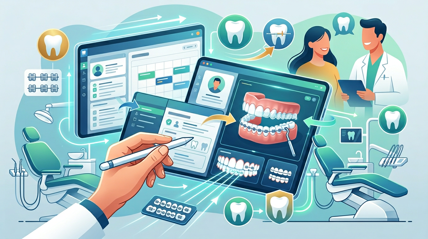
When integrated with dental software India practices use for scheduling and records — like Denti360 — digital imaging data sits alongside clinical notes, treatment plans, and billing in a single unified record. This integration removes redundant data entry and makes every patient's history immediately accessible at the point of care.
Practical Considerations for Indian Clinics Upgrading to Digital
The initial investment in digital imaging hardware is higher than maintaining an existing film setup. However, the medium-term economics typically favour digital. Ongoing costs for film, chemicals, darkroom maintenance, and proper chemical disposal are eliminated. Staff time saved in development and filing is redirected to patient care. And fewer missed early lesions mean lower rates of costly, avoidable restorative work — which benefits both patients and the clinic's reputation.
For clinics beginning the transition, PSP plates are often a practical first step: they are flexible like film, familiar in handling, and significantly less expensive than rigid CCD sensors. They require a dedicated scanner unit but otherwise slot into existing X-ray workflows with minimal disruption.
Conclusion
The evidence is clear: digital dental imaging outperforms film X-rays for early caries detection across every relevant metric — resolution, post-processing capability, radiation dose, workflow efficiency, and diagnostic consistency. For dental clinics in India committed to providing genuinely preventive care rather than simply managing disease after it progresses, upgrading to digital radiography is not a luxury — it is a standard of care decision.
Denti360 integrates with leading digital imaging systems to give your clinical data and administrative workflows a single home. Book a free Denti360 demo and see how the right dental practice management platform supports better diagnostics and smoother clinic operations from the ground up.