
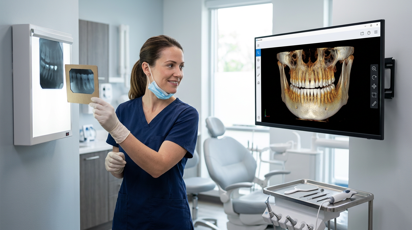
Accurate diagnosis is the foundation of excellent dental treatment. Everything downstream — the treatment plan, the materials selected, the prognosis given to the patient — depends on how well the clinician can see and interpret what is happening inside the tooth, below the gumline, and within the surrounding bone. For decades, two-dimensional film X-rays were the primary diagnostic window available to dental professionals. Today, digital imaging technology and sophisticated software platforms are fundamentally changing what dentists can see, how quickly they can see it, and how precisely they can act on that information. Denti360 dental imaging software sits at the centre of this transformation for Indian dental practices.
The Diagnostic Limitations That Led to a Technology Revolution
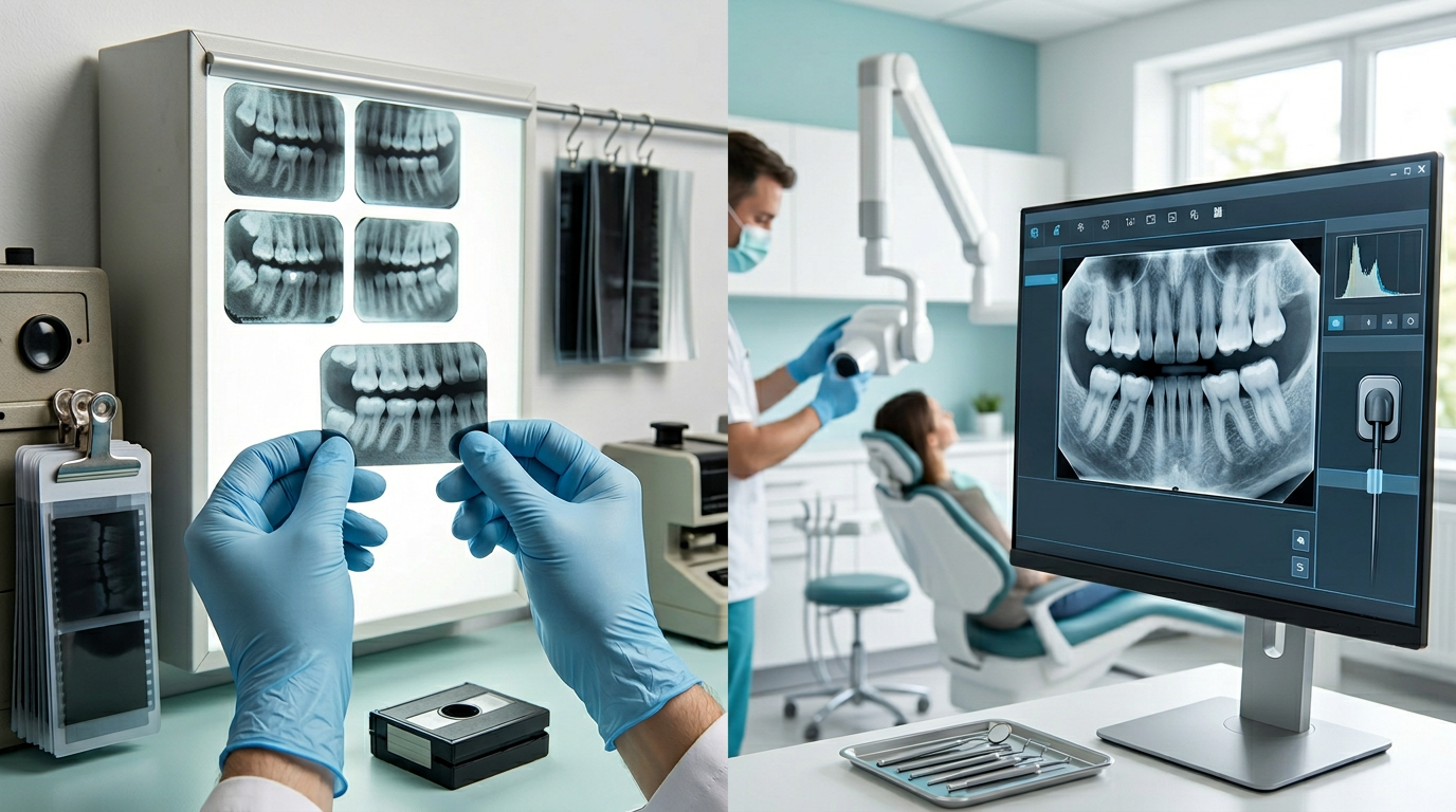
To appreciate what modern dental imaging software delivers, it helps to understand what clinicians were working with before. Traditional film radiography produces a two-dimensional shadow image of three-dimensional anatomy. This means that overlapping structures — adjacent roots, intersecting bone trabeculation, superimposed anatomical landmarks — can obscure exactly the detail that matters most. A periapical lesion hidden behind a dense cortical plate, a root fracture running in a direction perpendicular to the beam, an impacted third molar's relationship to the inferior alveolar canal: these are the diagnostic questions that two-dimensional film answers poorly.
Beyond image quality, the workflow of film radiography created practical constraints. Chemical processing introduced time delays and quality variables. Images could not be adjusted after development, meaning a slightly incorrect exposure often required a retake — and additional radiation for the patient. Physical storage of film archives created growing administrative burdens, and sharing images with specialists or insurers required physical handling that was slow and loss-prone.
These are not minor inconveniences. They translate directly into diagnostic uncertainty, treatment planning errors, and clinical risk. The development of digital radiography and, subsequently, cone beam computed tomography (CBCT) addressed many of these limitations. Denti360's imaging software goes further, providing an integrated environment in which all imaging data — from routine bitewings to three-dimensional volumetric scans — is managed, enhanced, and applied within a coherent clinical workflow.
What Denti360 Dental Imaging Software Actually Does
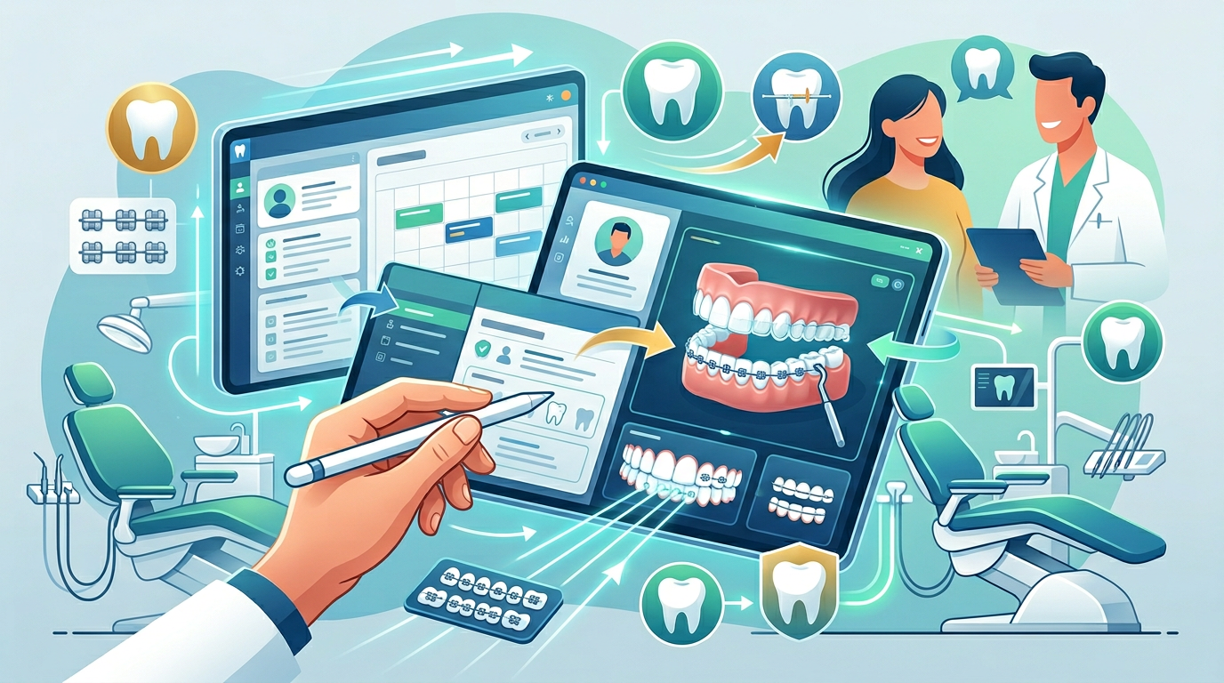
Denti360 is not simply a viewer for digital X-ray files. It is a diagnostic platform that integrates with the broader Denti360 dental practice management system, connecting imaging data with clinical notes, treatment plans, patient histories, and appointment records in a single unified interface.
Within the imaging module, clinicians have access to a range of tools that would have been impossible with film:
- Image enhancement controls: Brightness, contrast, gamma, and sharpness can be adjusted in real time, allowing the clinician to optimise the image for the specific diagnostic question at hand. Enhancement filters designed for caries detection or periodontal bone assessment highlight relevant structures without altering the underlying data.
- Precise measurement tools: Calibrated measurement tools allow clinicians to assess bone levels, root lengths, angulations, and anatomical distances directly on the image. For implant planning and orthodontic cephalometric analysis, this precision is clinically essential.
- Annotation and reporting: Clinicians can annotate images with text, arrows, and highlights — useful both for internal documentation and for patient education. Annotated images can be included in referral letters or printed for the patient to take home.
- Longitudinal comparison: Side-by-side display of current and previous radiographs makes it straightforward to monitor the progression of conditions over time — a capability that is particularly valuable for managing periodontal disease, watching a suspicious lesion, or evaluating orthodontic treatment progress.
CBCT Integration and Three-Dimensional Diagnostic Confidence
For cases requiring a three-dimensional perspective, Denti360 supports integration with CBCT scanners. The ability to view volumetric data — rotating the three-dimensional dataset to examine structures from any angle, isolating specific cross-sections, or generating coronal, sagittal, and axial views simultaneously — changes the diagnostic conversation in implantology, oral surgery, orthodontics, and complex endodontics.
Consider implant planning. With a two-dimensional panoramic film, the clinician can estimate available bone height but has limited information about width, density, and the precise three-dimensional relationship of the proposed implant site to adjacent roots, the sinus floor, or the inferior alveolar nerve. A CBCT scan, viewed and measured within Denti360, provides all of this information with a level of precision that makes the difference between a confident, well-planned procedure and one executed with unnecessary uncertainty.
In endodontics, three-dimensional imaging can reveal a missed canal, identify the source of persistent periapical pathology, or clarify the extent of a root fracture — diagnostic questions that two-dimensional films regularly leave unanswered. Denti360's support for CBCT data within the same imaging environment means that these advanced diagnostic capabilities are accessible without switching between disconnected software systems.
Radiation Safety and Patient Communication
Patients in India are increasingly informed about the healthcare choices they make, and questions about radiation exposure during dental X-rays are common. Denti360 supports the ALARA principle — As Low As Reasonably Achievable — in two practical ways. First, the software's compatibility with high-sensitivity digital sensors means that diagnostic-quality images can be produced at significantly lower radiation doses than traditional film required. Second, the post-processing tools available in Denti360 often allow a slightly sub-optimal initial exposure to be corrected digitally, rather than requiring a retake.
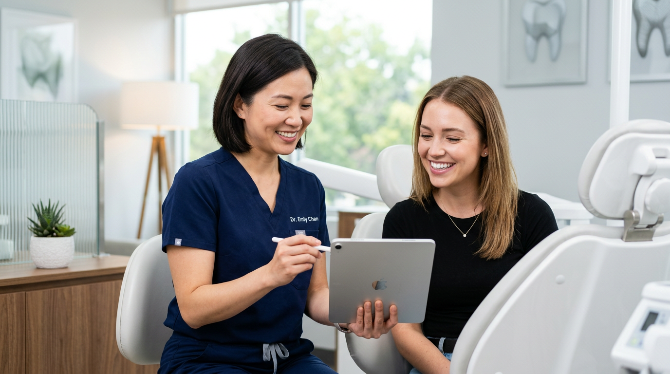
The ability to display large, high-resolution images on a monitor — and to annotate and explain them in real time — also transforms patient communication. When a patient can see a colour-highlighted image of their own bone loss, or a three-dimensional rendering of the space where an implant will be placed, the clinical conversation becomes more productive. Patients who understand their condition are more likely to accept recommended treatment and more likely to adhere to preventive recommendations. This is not a secondary benefit — it is a direct contribution to clinical outcomes.
Integration with Dental Practice Management for a Unified Workflow
One of the most practical advantages of Denti360's imaging software is its integration with the broader practice management platform. In practices where imaging is handled by a separate, disconnected system, clinicians and staff must toggle between applications, manually match images to patient records, and maintain two separate archives. This creates administrative friction, introduces opportunities for misallocation, and slows down both clinical and administrative workflows.
Within Denti360, images are automatically linked to the correct patient record at the time of capture. The clinician reviewing a patient's treatment plan can access their full radiographic history in the same interface, alongside notes from previous appointments, outstanding treatment items, and billing status. This unified view supports better-informed clinical decisions and a more coherent patient experience.
Conclusion: Precision Imaging as a Practice Standard
Diagnostic precision is not a luxury in modern dentistry — it is a standard that patients expect and clinical outcomes require. Denti360 dental imaging software gives Indian dental practices the tools to meet that standard consistently: high-resolution digital imaging, three-dimensional CBCT capability, powerful post-processing and measurement tools, and seamless integration with dental practice management workflows. The result is faster diagnosis, more confident treatment planning, better patient communication, and a practice environment that reflects the level of professionalism that distinguishes leading clinics.
If your practice is ready to upgrade its diagnostic capabilities and experience the difference that integrated imaging software makes, book a free Denti360 demo today. Speak with a specialist who understands the needs of Indian dental practices and see exactly how Denti360 can elevate your standard of care.