
Walk into any progressive dental clinic in India today and you are unlikely to find a darkroom. Digital X-rays have steadily replaced traditional film radiography across the country, and for good reason — the clinical, operational, and patient-experience advantages are difficult to argue with. Yet many practices still have questions about the transition: What exactly is the difference? What does a modern dental imaging setup actually deliver? And how does a platform like Denti360 fit into the picture? This article addresses those questions plainly, for dentists and clinic owners who want to make an informed decision.
How Traditional Film Radiography Works — and Where It Falls Short
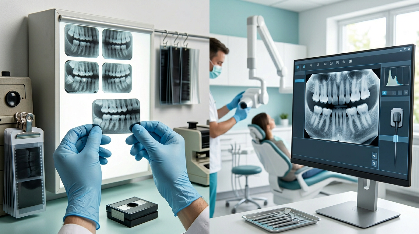
Film-based X-rays have served dentistry faithfully for over a century. The process is straightforward: ionising radiation passes through oral structures, exposing a silver-halide film that is then chemically developed in a darkroom. The resulting image reveals teeth, bone, and soft tissue in varying shades of grey, depending on tissue density.
The method works — but it comes with a list of limitations that become harder to ignore as better alternatives exist. Chemical processing takes time, introduces the risk of developing errors, and creates hazardous waste that must be disposed of properly. Film has a fixed dynamic range, meaning that a slightly over- or under-exposed image cannot be corrected after the fact — a retake is the only option, which means additional radiation exposure for the patient. Physical films need to be stored, organised, and retrieved manually, creating administrative overhead that grows with every passing year. And sharing a film X-ray with a specialist or insurer involves either posting a physical copy or scanning it — neither of which is fast or convenient.
What Digital Radiography Actually Delivers
Digital dental X-rays replace film with an electronic sensor or a reusable phosphor plate. When X-rays strike the sensor, the data is captured digitally and transmitted to a computer within seconds. The image appears on screen almost immediately — no developing, no chemicals, no waiting.
The practical benefits flow directly from this architecture:
- Significantly reduced radiation exposure: Digital sensors are far more sensitive to X-rays than film, which means the exposure dose needed to produce a diagnostic image is considerably lower — often reduced by 70 percent or more compared to conventional film. This matters for patients who require regular monitoring, and it matters for the ALARA principle that guides safe radiographic practice.
- Instant image availability: Images appear on screen within seconds of exposure. For a busy clinic, this alone has a tangible impact on appointment flow — no waiting for processing, no gaps in the consultation while film develops.
- Post-processing flexibility: Digital images can be adjusted for brightness, contrast, and magnification after capture. A slightly under-exposed image that would have required a retake on film can often be corrected digitally, reducing unnecessary radiation exposure and saving appointment time.
- Better diagnostic detail: Modern digital sensors offer high resolution and a wide dynamic range, making it easier to detect early caries, subtle bone changes, and fine root anatomy that might be missed on film.
- Easy storage, retrieval, and sharing: Digital images are stored in the practice management system and retrievable instantly by patient name or date. Sending images to a specialist or insurer takes seconds rather than days.
The Denti360 Advantage in Dental Imaging
Having digital hardware is only part of the equation. The software that manages, displays, and integrates those images into the broader patient record is equally important — and this is where Denti360 provides a meaningful edge for Indian dental practices.
Denti360's dental imaging software is built to work within a complete dental practice management workflow rather than as a standalone imaging viewer. When a patient's X-rays are captured, they are automatically linked to that patient's record, alongside their clinical notes, treatment plan, and appointment history. There is no separate login, no manual file import, and no risk of images being associated with the wrong patient.
Within the imaging module, clinicians can use measurement tools to assess bone levels and tooth dimensions, apply enhancement filters to highlight specific structures, and annotate images for patient education or referral letters. Side-by-side comparison of current and historical radiographs makes it easy to track the progression of conditions like periodontal bone loss or a developing carious lesion — a capability that strengthens both clinical decision-making and patient communication.
For practices that use cone beam computed tomography (CBCT) for implant planning, orthodontic assessment, or complex endodontic cases, Denti360 supports 3D volumetric imaging within the same integrated environment. The ability to view true three-dimensional anatomy — without superimposition of overlapping structures — represents a significant leap in diagnostic confidence for these applications.
Patient Safety, Data Security, and Compliance
Two concerns that come up frequently in discussions about digital imaging are data security and radiation safety. Denti360 addresses both directly.
On the radiation side, the lower exposure requirements of digital sensors mean that patients receive meaningfully less ionising radiation over the course of their dental care compared to film-based practices. Denti360's exposure settings are configurable to patient size and the clinical requirement, helping practices apply the minimum dose needed to achieve a diagnostic image.
On data security, all images stored within Denti360 are encrypted and access-controlled. Role-based permissions ensure that staff members can only view the records relevant to their function. Automated backups protect against data loss, and the system's architecture is designed to support compliance with Indian healthcare data regulations as they continue to evolve. Physical films, by contrast, can be damaged, misfiled, or accessed by anyone who can locate the folder — a significant vulnerability that digital storage eliminates.
Making the Transition: What to Expect
Practices considering the move from film to digital imaging sometimes worry about disruption during the changeover. In practice, the transition is more straightforward than expected, particularly with the right implementation support. Denti360's onboarding process includes hardware setup and calibration, integration with your existing patient management workflows, and hands-on training for clinical and administrative staff. Most practices find that their team is comfortable with the new system within a few weeks, and the operational benefits — faster appointments, easier image retrieval, no darkroom maintenance — become apparent almost immediately.
Conclusion: Modern Dentistry Deserves Modern Imaging
The case for digital X-rays over traditional film is not a matter of preference — it is a matter of clinical quality, patient safety, and operational efficiency. Lower radiation exposure, instant image availability, superior diagnostic detail, and seamless integration with dental practice management systems make digital radiography the clear standard for contemporary dental care in India.
Denti360 brings all of these advantages together in a platform built specifically for the needs of Indian dental clinics — from the single-chair general practice to the multi-specialty centre. If you are ready to upgrade your imaging capabilities and see how Denti360 can transform the diagnostic workflow in your practice, book a free Denti360 demo today and speak with a specialist who understands the Indian dental landscape.