
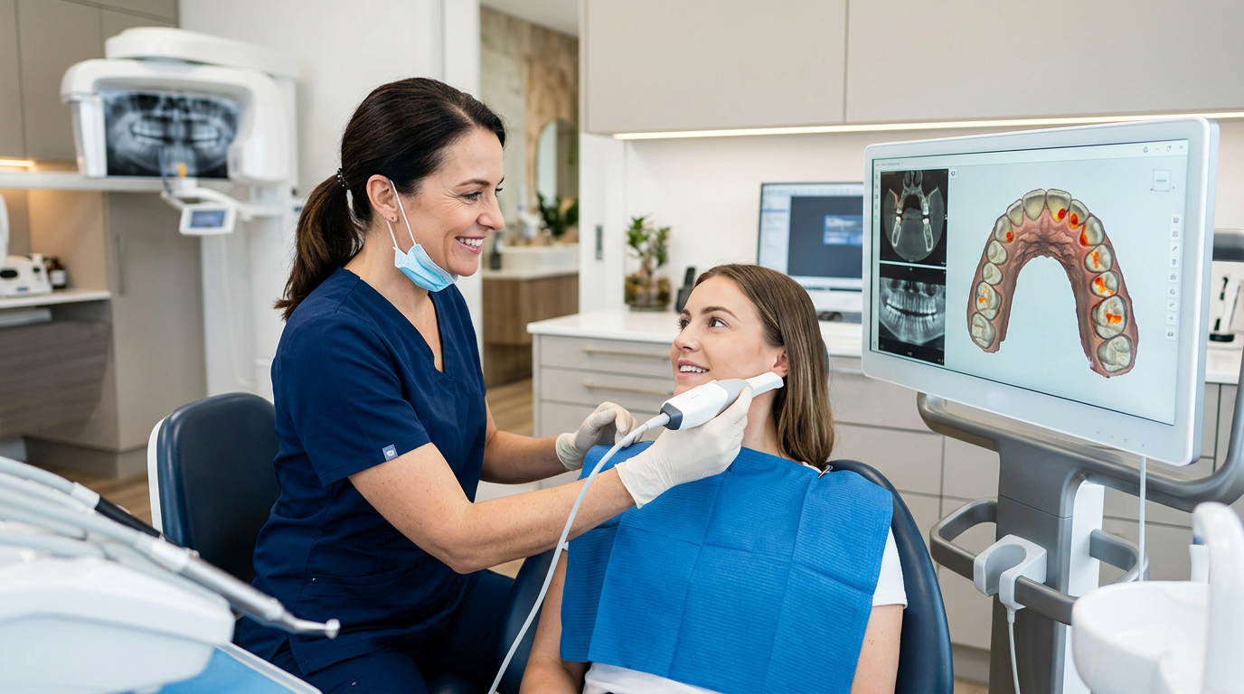
5 Ways Integrated Dental Imaging Improves Caries Detection Accuracy
Integrated dental imaging is changing how practices catch tooth decay before it becomes a costly problem, and that matters because caries remains one of the most prevalent chronic diseases globally, yet incipient lesions, interproximal caries, and secondary caries under restorations routinely slip past traditional methods. Early, accurate detection is paramount to effective preventive care, minimally invasive treatment, and preserving long-term oral health. For dental professionals at Denti360 and beyond, leveraging these advanced systems is not just an option but a necessity in providing the highest standard of patient care.
Integrated dental imaging refers to the seamless combination and often synergistic use of various imaging modalities within a unified digital workflow. This approach moves beyond isolated techniques, creating a comprehensive visual record that aids more precise detection, diagnosis, treatment planning, and monitoring. By combining different perspectives and data points, practitioners gain a multi-dimensional understanding of a patient's oral health, significantly improving their ability to detect caries earlier and with greater confidence. This post explores five transformative ways integrated dental imaging can improve caries detection accuracy.
1. The Synergistic Power of Digital Radiography and Intraoral Cameras
The foundational pillars of digital dental imaging, digital radiography (DR) and intraoral cameras (IOCs), offer a powerful synergistic approach to caries detection when integrated. Digital radiography provides invaluable insight into subsurface tooth structures, revealing demineralization in enamel and dentin that is often invisible to the naked eye. With significantly reduced radiation exposure compared to film-based radiography, DR images offer superior clarity, contrast, and the ability to be manipulated for enhanced visualization. Features like zoom, contrast adjustment, and measurement tools let clinicians scrutinize suspicious areas with high precision.
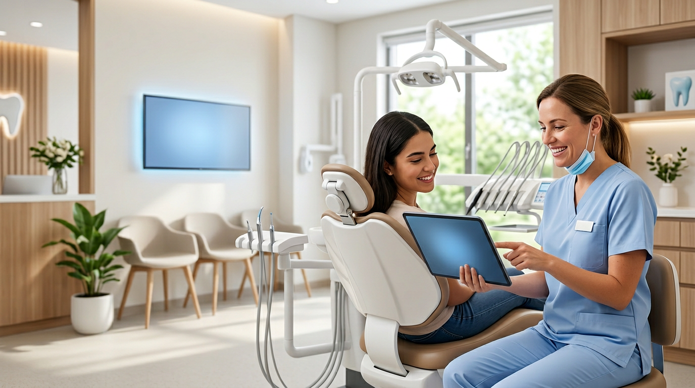
However, DR has limitations. It is a 2D representation of a 3D structure and can suffer from superimposition, making early occlusal or buccal/lingual caries detection challenging. This is where intraoral cameras become indispensable. IOCs provide real-time, magnified, full-color documentation of the tooth surface. They excel at identifying changes in surface texture, color alterations, and visible signs of demineralization, particularly on occlusal surfaces, pits, fissures, and around existing restorations. Showing patients their own teeth in high definition aids diagnosis and significantly enhances patient education and treatment acceptance.
When these two technologies are integrated, the diagnostic power multiplies. A suspicious area identified on a bitewing radiograph can be cross-referenced with a high-resolution intraoral image of the corresponding surface. Conversely, a visually questionable spot observed with the IOC can prompt a targeted radiographic assessment. The Denti360 platform, for instance, allows quick toggling between radiographic and intraoral views, helping dentists piece together a more complete diagnostic picture. This integrated workflow reduces diagnostic uncertainty, minimizes the chance of missing caries, and prevents unnecessary restorative interventions by allowing a more conservative, watchful-waiting approach when appropriate. Furthermore, sequential images from both modalities over time provide an objective record for monitoring lesion progression or arrest, crucial for personalized preventive strategies.
2. Advanced Fluorescence-Based Caries Detection Systems
Beyond traditional visual and radiographic methods, fluorescence-based caries detection systems represent a significant leap forward, offering a non-invasive way to identify demineralized tooth structure and bacterial activity. These systems, such as DIAGNOdent or similar technologies, operate on the principle that healthy enamel exhibits natural fluorescence when exposed to specific wavelengths of light. Caries lesions, due to demineralization and the presence of porphyrins produced by cariogenic bacteria, absorb and scatter light differently, leading to a loss of fluorescence or an emission of red fluorescence depending on the technology.
Integrating these systems into the imaging workflow provides an objective, quantifiable measurement of demineralization, particularly valuable for detecting occlusal and smooth-surface caries that may be difficult to discern radiographically or visually. For example, a clinician might perform an initial visual inspection with an intraoral camera, followed by a radiographic assessment. If ambiguity remains, or if a high-risk patient presents, the fluorescence-based system can provide a numerical reading or color-coded map indicating the presence and extent of caries. This objective data helps differentiate between stain and actual carious lesions, reducing false positives and false negatives.
The Denti360 platform can integrate data from these fluorescence systems, displaying the readings alongside radiographic and intraoral images. This comprehensive view allows a more confident diagnosis, especially for incipient lesions that may be candidates for remineralization therapy rather than immediate restoration. By providing quantitative data, these systems also enable precise monitoring of lesions over time, assessing the effectiveness of preventive measures. This proactive approach supports minimally invasive dentistry by facilitating intervention at the earliest stages, often before cavitation occurs. The ability to objectively demonstrate caries to patients using these visual and numerical aids also significantly improves patient understanding and treatment acceptance, fostering a more collaborative approach to oral health management.
3. Cone Beam Computed Tomography (CBCT) for Complex Cases
While digital radiography is excellent for routine caries detection, its 2D nature can sometimes limit the ability to accurately assess the depth and extent of complex or overlapping lesions. For specific challenging cases, integrating Cone Beam Computed Tomography (CBCT) into the diagnostic protocol can dramatically improve caries detection accuracy. CBCT provides high-resolution, three-dimensional images of the dentition and surrounding bone structures, offering unparalleled anatomical detail.
Although CBCT involves a higher radiation dose than conventional digital radiographs, its use is justified when 2D images are inconclusive or when precise 3D information is critical for treatment planning. For caries detection, CBCT becomes invaluable in situations such as:
- Deep or Extensive Caries: Accurately assessing a lesion's proximity to the pulp chamber, crucial for determining restorative vs. endodontic treatment.
- Secondary Caries Around Complex Restorations: Identifying recurrent decay under large crowns, onlays, or extensive composite fillings where radiographic superimposition is a significant challenge.
- Cracked Teeth Syndrome: Differentiating between non-carious cracks and those associated with early carious demineralization.
- Interproximal Caries in Crowded Dentition: Overcoming superimposition issues in crowded anterior or posterior segments.
- Identifying Root Caries: Especially in areas where soft tissue or bone obscures the view.
The Denti360 imaging software allows seamless integration of CBCT scans with other modalities. A clinician can view a 3D reconstruction of a tooth alongside its 2D digital radiograph and intraoral camera images. This multi-modal approach provides a holistic view, allowing precise localization, accurate measurement of lesion size, and detailed assessment of its relationship to adjacent structures. This level of detail minimizes diagnostic guesswork, leading to more predictable treatment outcomes and reducing the need for exploratory procedures. Knowing the exact extent of decay pre-operatively lets dentists plan more conservatively, preserving healthy tooth structure and minimizing patient discomfort.
4. AI-Powered Caries Detection and Diagnostic Support
The most transformative frontier in integrated dental imaging is the incorporation of artificial intelligence (AI) and machine learning algorithms for caries detection. AI-powered software tools analyze vast amounts of radiographic and intraoral image data, learning to identify patterns associated with caries that might be subtle or easily missed by the human eye, even an experienced one.
Integrated AI solutions within platforms like Denti360 can:
- Automate Caries Localization: AI algorithms quickly scan digital radiographs and intraoral images, highlighting suspicious areas and potential carious lesions for the clinician's review. This acts as a 'second pair of eyes,' reducing fatigue and improving consistency.
- Quantify Lesion Progression: Over successive appointments, AI can compare images to objectively measure changes in lesion size or density, providing quantitative data on whether a lesion is progressing, arresting, or remineralizing. This is invaluable for guiding treatment decisions, especially watchful-waiting strategies.
- Enhance Image Interpretation: AI can be trained to recognize early demineralization patterns or secondary caries obscured by restorations, providing prompts or confidence scores to assist diagnosis.
- Reduce Inter-Operator Variability: By providing a standardized analysis framework, AI helps reduce discrepancies in diagnosis between clinicians, leading to more consistent patient care across a practice or network.
It's crucial to emphasize that AI in caries detection is a diagnostic support tool, not a replacement for clinical judgment. The dentist remains the ultimate decision-maker. However, by offloading the laborious task of initial screening and pattern recognition, AI frees the clinician to focus on complex cases, patient communication, and comprehensive treatment planning. Integrating AI into the existing imaging workflow means that as new radiographs or intraoral photos are captured, they are instantly analyzed, providing real-time diagnostic assistance. This enhances accuracy and improves workflow efficiency and patient throughput, ensuring no potential lesion goes unnoticed in a busy practice. As AI technology continues to evolve, its role in refining caries detection and preventive strategies is poised to become even more profound by 2025 and beyond.
5. Seamless Integration With Practice Management and EHR Systems
While the previous points focused on individual modalities and AI, the ultimate improvement in caries detection accuracy comes from seamlessly integrating all these systems with the practice's overarching Practice Management Software (PMS) and Electronic Health Records (EHR) system. An integrated Denti360 ecosystem means all patient data, clinical notes, treatment history, and crucially all imaging data from radiographs, intraoral cameras, fluorescence systems, and CBCT scans, resides in a unified, easily accessible digital file. This kind of streamlined patient management keeps every diagnostic detail in one place.
This holistic integration profoundly impacts caries detection in several ways:
- Comprehensive Patient History: Clinicians can instantly access historical images and diagnostic notes, allowing longitudinal comparison of suspicious areas. Tracking lesion progression or regression over months or years with a complete visual and textual record is vital for accurate diagnosis and tailored management plans.
- Streamlined Workflow: Imaging data is automatically linked to the correct patient and tooth, eliminating manual data entry, reducing errors, and saving valuable chair time. This efficiency dedicates more time to patient examination and treatment.
- Enhanced Collaboration: Integrated systems facilitate easy sharing of images and reports with specialists for second opinions or complex treatment planning, ensuring a multidisciplinary approach to challenging caries cases.
- Improved Patient Education: Displaying various images (radiographs, intraoral photos, fluorescence readings) side-by-side on a monitor lets dentists more effectively educate patients about their oral condition, the presence of caries, and the proposed treatment. This transparency builds trust and improves treatment acceptance.
- Quality Assurance and Auditability: A fully integrated system provides a robust audit trail, documenting every image captured, diagnosis made, and treatment rendered, crucial for quality assurance, compliance, and even forensic purposes.
For a Denti360 practice, this means that from the moment a patient sits in the chair, their complete oral health story, meticulously documented through various imaging and clinical data, is at the dentist's fingertips. This comprehensive overview ensures all relevant information is considered during caries detection, leading to more accurate diagnoses, appropriate treatment decisions, and ultimately better patient outcomes. The ability to pull up previous images and compare them side-by-side with current ones is an unparalleled diagnostic advantage, letting dentists confidently identify even subtle changes that might indicate early caries.
Conclusion: The Future of Caries Detection Is Integrated
The journey toward improving caries detection accuracy is continuous, driven by technological innovation and a commitment to superior patient care. The five strategies outlined, the synergistic use of digital radiography and intraoral cameras, advanced fluorescence-based detection, the precision of CBCT for complex cases, the revolutionary support of AI, and seamless integration with practice management systems, collectively represent the cutting edge of modern dental diagnostics. By embracing these integrated imaging solutions, dental professionals at Denti360 and across the globe are empowered to detect caries earlier, diagnose with greater confidence, treat more conservatively, and safeguard the oral health of their communities more effectively than ever.
Investing in and intelligently implementing these integrated systems is not just a technology upgrade; it's an investment in diagnostic excellence, operational efficiency, and enhanced patient trust. As we move into 2025 and beyond, the practice that leverages these powerful tools will be best positioned to deliver the highest standard of preventive and restorative dentistry. To see how an integrated imaging and records ecosystem fits your workflow, explore the Denti360 platform features or request a demo and solidify your role as a leader in comprehensive oral healthcare.