
Dental Imaging Software vs. Traditional X-rays: Interproximal Cavity Clarity
Dental imaging software is changing how clinicians spot the interproximal cavities hiding between teeth, where traditional film so often falls short and early lesions slip past undetected. Early, accurate caries detection is crucial for timely intervention and preserving oral health, yet conventional film-based X-rays carry real diagnostic limits. As digital technology advances, dental imaging software paired with digital radiography has emerged as a formidable contender. This post compares dental imaging software against traditional X-rays, evaluating which delivers superior clarity for identifying interproximal cavities. Denti360 is committed to insights on the technologies shaping dental care, equipping practitioners to make informed decisions for their practices and patients.
Understanding Traditional X-rays: The Foundation of Dental Diagnostics
Traditional film-based X-rays, also known as conventional radiography, have anchored dental diagnostics for over a century. The process exposes a dental film to a controlled burst of X-rays. As they pass through oral structures, different tissues absorb varying amounts of radiation, creating a latent image on the film that is then developed using chemical solutions in a darkroom. The resulting radiograph provides a two-dimensional view of teeth, bone, and surrounding structures.
Mechanism and Image Formation
Traditional X-rays rely on differential absorption by various tissues. Denser structures like enamel and bone absorb more X-rays and appear radiopaque (white or light grey). Less dense structures, such as dental pulp or soft tissue, let more X-rays pass and appear radiolucent (darker grey or black). Cavities, being demineralized and less dense than healthy tooth structure, appear as radiolucencies on the film.
Advantages of Traditional X-rays
- Established and Familiar: Dentists are trained on and highly familiar with interpreting traditional radiographs.
- Cost-Effective Initial Setup: The initial investment for traditional X-ray equipment can sometimes be lower than for digital systems.
- Universally Understood: The diagnostic principles are well understood across the dental community.
Limitations of Traditional X-rays for Interproximal Cavities
Despite their long-standing utility, traditional X-rays present several limitations for the nuanced detection of interproximal cavities:
- Limited Dynamic Range: Film has a relatively narrow dynamic range, struggling to capture a wide spectrum of tissue densities in one exposure. This can underexpose or overexpose areas, obscuring subtle demineralization that signals early interproximal caries.
- Chemical Processing Variability: Final image quality depends heavily on chemical processing. Variations in solution temperature, development time, and chemical freshness affect clarity, causing artifacts, fogging, or reduced contrast that make subtle interproximal lesions harder to discern.
- Geometric Distortion: The dental arch's curvature and X-ray cone angulation cause geometric distortion and superimposition of structures, masking interproximal cavities, especially small ones or those in overlapping areas.
- Time-Consuming Workflow: Exposing, developing, and mounting film is time-consuming, prolonging appointments and reducing efficiency.
- Environmental Concerns: Film-processing chemicals are hazardous and require proper disposal, posing environmental challenges.
- Lack of Post-Processing Capabilities: Once developed, a traditional film image cannot be digitally enhanced or manipulated for improved clarity.
Embracing Dental Imaging Software with Digital X-rays: A New Era of Precision
Digital radiography paired with sophisticated dental imaging software represents a paradigm shift in diagnostics. Instead of film, digital sensors or phosphor plates capture X-ray images, converting X-ray photons directly into an electrical signal that is digitized and sent to a computer. The dental imaging software then processes, displays, and stores these images. See the features that modern dental software brings to a practice.
Mechanism and Image Formation
Digital X-ray systems use CCD (Charge-Coupled Device), CMOS (Complementary Metal-Oxide-Semiconductor) sensors, or PSP (Photostimulable Phosphor) plates. Both capture X-ray data and convert it into digital information, rendered into an image viewable instantly on a monitor. The key advantage is direct digital output that software can manipulate and enhance.
Core Components of Digital Imaging Systems
- Digital Sensors/PSP Plates: Replace traditional film, capturing X-ray data electronically.
- X-ray Unit: Emits the X-rays, often with lower radiation doses than traditional units.
- Computer: Houses the dental imaging software and displays the images.
- Dental Imaging Software: The brain of the system, enabling image acquisition, processing, enhancement, and management.
Advantages of Dental Imaging Software for Interproximal Cavities
The true power of digital radiography for subtle lesions like interproximal cavities lies in its software. Here's why it often delivers superior clarity:
- Enhanced Image Quality and Clarity:
- Higher Resolution and Contrast: Digital sensors generally offer a wider dynamic range and superior contrast resolution than film, capturing more subtle density differences and making early enamel demineralization, characteristic of interproximal cavities, more discernible.
- Instantaneous Image Acquisition: Images appear on screen within seconds, allowing immediate review and adjustment, optimizing diagnostic quality during the visit.
- Powerful Post-Processing Capabilities: This is where dental imaging software truly shines for interproximal cavity detection:
- Brightness and Contrast Adjustment: Dentists dynamically adjust brightness and contrast after acquisition to fine-tune the image and reveal subtle radiolucencies hard to see on static film. For interproximal areas, this means lightening or darkening regions to better visualize early enamel demineralization.
- Magnification and Zoom: Dentists zoom in on areas of interest without pixelation for a magnified view of interproximal surfaces, examining suspicious areas in detail and differentiating true cavities from artifacts or healthy anatomy.
- Sharpening and Edge Enhancement Filters: These filters sharpen edges and enhance detail, making cavity boundaries more distinct against surrounding tooth structure, particularly beneficial for small or incipient interproximal lesions.
- Inversion (Negative Image): Some software inverts the image so radiolucent areas appear white and radiopaque areas black. For certain practitioners, this reversed contrast makes subtle interproximal cavities more apparent.
- Colorization and Pseudocolor: Though not universally used for interproximal detection, some advanced software color-maps different densities, sometimes highlighting demineralization in a visually distinct way.
- Measurement Tools: Software often includes tools to measure lesion size and depth, aiding treatment planning and monitoring progression over time.
- Reduced Radiation Exposure: Digital X-ray systems typically require significantly less radiation (up to 90% less) than traditional film, enhancing patient safety.
- Environmental Friendliness: Eliminates hazardous chemicals and film, contributing to a greener practice.
- Efficient Workflow and Storage: Images are stored digitally instantly, eliminating physical film filing and retrieval. This streamlines workflow and reduces administrative burden, with easy sharing to specialists or insurers as a further advantage.
- Patient Education: Displaying and manipulating images instantly in front of the patient is a powerful educational tool. Dentists zoom in on cavities, explain findings clearly, and demonstrate the need for treatment, fostering greater understanding and acceptance.
Direct Comparison: Interproximal Cavity Detection
For the crucial task of detecting interproximal cavities, the advantages of dental imaging software become strikingly clear.
Clarity and Diagnostic Accuracy
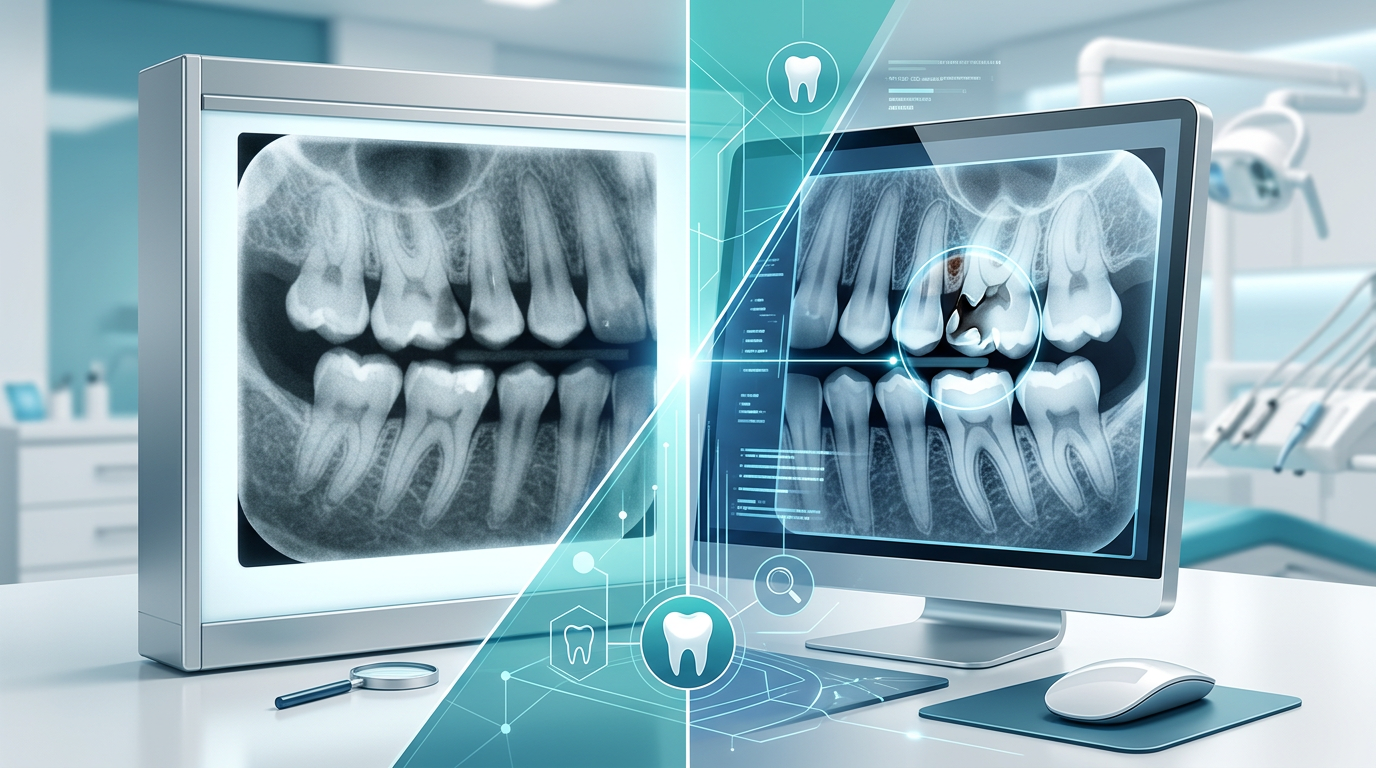
Traditional X-rays: Capable of detecting larger interproximal cavities, traditional film struggles with early or incipient lesions. Fixed contrast and resolution, plus potential processing errors and geometric limitations, can cause missed diagnoses or repeat exposures, delaying treatment and allowing caries to progress.
Dental Imaging Software: The ability to manipulate and enhance digital images significantly boosts clarity for interproximal cavities. Dentists fine-tune brightness and contrast to highlight subtle demineralization, zoom in for closer inspection, and apply sharpening filters to define lesion boundaries. This superior clarity enables earlier detection of incipient lesions still confined to enamel, allowing preventive interventions or less invasive restorative procedures. Consistent digital image quality, free from chemical processing variability, also raises diagnostic accuracy.
User Experience and Efficiency
Traditional X-rays: Manual film processing is labor-intensive, time-consuming, and prone to human error, with the darkroom environment and chemical handling adding complexity and cost.
Dental Imaging Software: Offers an intuitive, streamlined workflow. Images are available almost instantly, reducing chair time and increasing efficiency, while easy storage, retrieval, and sharing enhance collaboration and record-keeping. Discover the broader benefits of a streamlined practice.
Radiation Safety
Traditional X-rays: Requires higher radiation doses than digital systems.
Dental Imaging Software: Significantly reduces patient and operator radiation exposure, aligning with the ALARA (As Low As Reasonably Achievable) principle of radiation protection, a considerable advantage for patient safety and practice health standards in 2025.
Cost Implications Over Time
Traditional X-rays: Lower initial setup cost but ongoing expenses for film, chemicals, and disposal, plus darkroom equipment maintenance.
Dental Imaging Software: Higher initial investment in digital sensors/PSP plates and software licenses. However, long-term savings accrue from eliminating film and chemicals, reduced labor costs, and improved efficiency. Enhanced diagnostics also potentially lead to better outcomes and patient retention.
The Future of Dental Imaging: AI Integration and Beyond
The dental imaging landscape keeps evolving, and the software is a platform for ongoing innovation. Artificial intelligence (AI) is already integrated into some advanced imaging solutions. AI algorithms can help highlight potential areas of concern, such as interproximal cavities, by analyzing radiographic patterns and drawing attention to subtle changes the human eye might miss. While AI is a powerful diagnostic aid, it augments rather than replaces the expertise of the dental professional.
Further advances in 3D imaging, such as Cone Beam Computed Tomography (CBCT), offer unprecedented views of oral structures, though routine interproximal cavity detection is generally limited by higher radiation doses compared to 2D digital radiography. Still, the convergence of these technologies promises even greater diagnostic accuracy in the years leading up to 2025 and beyond.
The Clear Choice for Modern Dentistry
Traditional X-rays have a venerable history and still hold a place in some practices, but dental imaging software paired with digital radiography unequivocally offers superior clarity for detecting interproximal cavities. Its advanced post-processing, including brightness and contrast adjustment, magnification, and sharpening filters, lets dentists visualize subtle lesions with detail unattainable on film. This enhanced clarity translates into earlier diagnosis, more conservative treatment planning, improved patient safety through reduced radiation, and a more efficient, environmentally friendly practice.
For Denti360, advocating technologies that elevate diagnostic precision and patient care is fundamental. The transition to digital imaging software isn't merely an upgrade; it's an investment in the future of dental diagnostics, ensuring practitioners provide the highest standard of care and detect challenging lesions like interproximal cavities with unmatched clarity and confidence. Ready to position your practice at the forefront of modern oral healthcare? Connect with the Denti360 team to explore the technology that delivers optimal patient outcomes in 2025 and beyond.